. 2001 Jun;12(6):1725-36.
doi: 10.1091/mbc.12.6.1725.

Distribution and transport of cholesterol in Caenorhabditis elegans

Affiliations
Free PMC article

Distribution and transport of cholesterol in Caenorhabditis elegans

V Matyash et al. Mol Biol Cell. 2001 Jun.
Free PMC article

Abstract

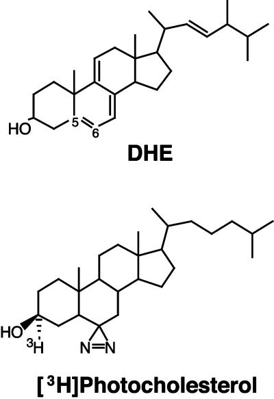
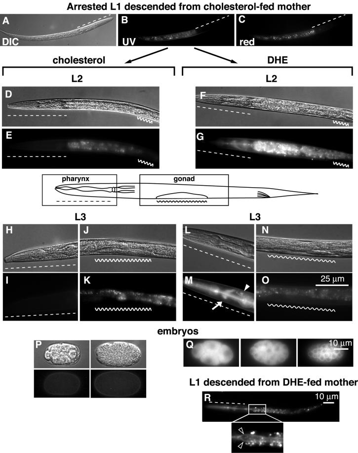
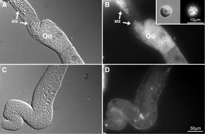
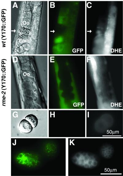
Cholesterol transport is an essential process in all multicellular organisms. In this study we applied two recently developed approaches to investigate the distribution and molecular mechanisms of cholesterol transport in Caenorhabditis elegans. The distribution of cholesterol in living worms was studied by imaging its fluorescent analog, dehydroergosterol, which we applied to the animals by feeding. Dehydroergosterol accumulates primarily in the pharynx, nerve ring, excretory gland cell, and gut of L1-L3 larvae. Later, the bulk of dehydroergosterol accumulates in oocytes and spermatozoa. Males display exceptionally strong labeling of spermatids, which suggests a possible role for cholesterol in sperm development. In a complementary approach, we used a photoactivatable cholesterol analog to identify cholesterol-binding proteins in C. elegans. Three major and several minor proteins were found specifically cross-linked to photocholesterol after UV irradiation. The major proteins were identified as vitellogenins. rme-2 mutants, which lack the vitellogenin receptor, fail to accumulate dehydroergosterol in oocytes and embryos and instead accumulate dehydroergosterol in the body cavity along with vitellogenin. Thus, uptake of cholesterol by C. elegans oocytes occurs via an endocytotic pathway involving yolk proteins. The pathway is a likely evolutionary ancestor of mammalian cholesterol transport.


Figures

Figure 1
Figure 1
Structures of DHE and [3H]photocholesterol.
Figure 2
Figure 2
Labeling of L1, L2, and L3 larval stages with DHE. (A–C) L1 larva descending from cholesterol-fed mother. L2 larva produced by the transfer of L1 onto cholesterol- (D and E) or DHE-containing (F and G) plate. Pharingeal (H and I) and gonadal (J and K) regions of L3 larva grown on control plate and correspondingly pharingeal (L and M) and gonadal (N and O) regions of DHE-fed worm. Different developmental stages of C. elegans embryos grown in control conditions (P) and on DHE plate (Q). DHE fluorescence distribution in L1 larvae descended from DHE-fed mother (R). Inset in R shows magnified region of gut. Bright spots around the apical surfaces of the gut are autofluorescent gut granules. Dashed line, pharyngeal region; wavy line, gonad; arrow, nerve ring; closed arrowhead, excretory gland cell; open arrowheads, apical surfaces of the gut.
Figure 3
Figure 3
Labeling of late L4 larvae (A–D), young adult hermaphrodite (E–H), adult hermaphrodite (I–L), and adult virgin male (M–P) with DHE. (A, B, E, F, I, J, M, and N) Gonadal regions of worms grown on cholesterol and (C, D, G, H, K, L, O, P) on DHE plates. std, spermatids; stz, spermatozoa; arrowhead, proximal spermathecal valve.
Figure 4
Figure 4
Hermaphrodite and male sperm from dissected animals. (A and B) Spermatheca and proximal gonad of a dissected hermaphrodite; (C and D) distal gonad of the same animal. Inset in B displays a spermatid dissected from him-8 male grown on DHE plate. Oo, oocyte; stz, spermatozoa.
Figure 5
Figure 5
Labeling of mixed population (MP) and L4/young adults with [3H]photocholesterol (A). Major bands of high-molecular mass are indicated with arrowheads and minor bands with dots. The separation was performed on a 12% acrylamide gel. Lysates from UV-irradiated (+) and from nonirradiated (−) worms. (B) Separation of labeled bands from mixed population on a 5% gel. (C) Extraction of labeled proteins with Triton X-114 and subsequent phase separation. AP, aqueous phase; W1 and W2, washes 1 and 2, respectively; DP, detergent phase. (D) Comparison of the labeling in adult hermaphrodites and males. (E) Immunoprecipitation of cross-linked bands with nonimmune mouse IgG, polyclonal anti-YP170 and monoclonal antibodies PIIA3 and OIC1. (F) Labeling of YP170::gfp strain with [3H]photocholesterol. wt, lysate from wild-type worms.
Figure 6
Figure 6
Uptake of DHE in rme-2 strain. Adult hermaphrodite of vit-2::gfp strain, grown on DHE plate, observed with the use of DIC optics (A) or in the GFP (B) and DHE (C) fluorescence channels. rme-2 mutant expressing YP170::GFP was fed with DHE and observed in DIC (D), GFP (E), and DHE (F) channels. Oocytes from rme-2 mutant expressing YP170::GFP grown on DHE imaged with the use of DIC (G), GFP (H) and DHE (I) channels. Embryos from YP170:: GFP strain grown on DHE, observed in GFP (J) and DHE (K) channels. Green, GFP fluorescence; arrow, spermatozoa; Oo, oocyte; Emb, embryo.
Figure 7
Figure 7
Separation of vitellogenin complexes from rme-2 mutants by centrifugation on a sucrose gradient. The graphs with closed squares and circles depict respectively total radioactivity and radioactivity after TCA-precipitation in the fractions from rme-2 extract. □, total radioactivity; ○, radioactivity in TCA-precipitated fractions of the extract from wild-type worms. Inset: labeled bands from TCA-precipitated fractions 10 and 13 of rme-2 extract, separated on a 12% SDS-PAGE.

References

    1. Austin CR. The 'capacitation' of the mammalian sperm. Nature. 1952;170:326. - PubMed
    1. Baker ME. Is vitellogenin an ancestor of apolipoprotein B-100 of human low-density lipoprotein and human lipoprotein lipase? Biochem J. 1988;255:1057–1060. - PMC - PubMed
    1. Bargmann CI, Mori I . a.C.o.C.e. Researchers, editors. Chemotaxis and thermotaxis. In: Wood WB, editor. The Nematode Caenorhabditis elegans. Cold Spring Harbor, NY: Cold Spring Harbor Laboratory Press; 1988. pp. 717–737.
    1. Bottjer KP, Weinstein PP, Thompson MJ. Effects of an azasteroid on growth, development and reproduction of the free-living nematodes Caenorhabditis briggsae and Panagrellus redivivus. Comp Biochem Physiol. 1985;82B:99–106. - PubMed
    1. Brenner S. The genetics of Caenorhabditis elegans. Genetics. 1974;77:71–94. - PMC - PubMed

Publication types

MeSH terms