. 2001 Jun;12(6):1751-64.
doi: 10.1091/mbc.12.6.1751.

Spindle dynamics and the role of gamma-tubulin in early Caenorhabditis elegans embryos

Affiliations
Free PMC article

Spindle dynamics and the role of gamma-tubulin in early Caenorhabditis elegans embryos

S Strome et al. Mol Biol Cell. 2001 Jun.
Free PMC article

Abstract

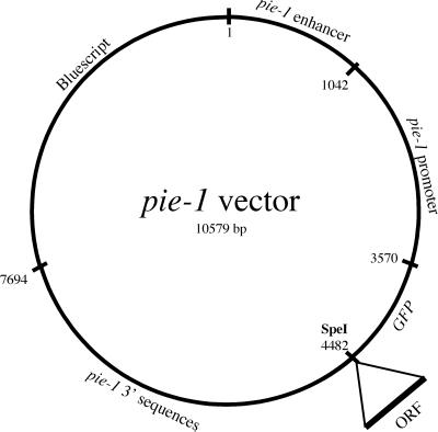
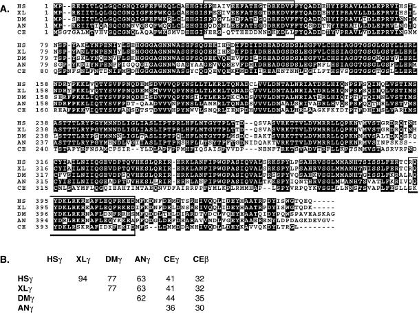
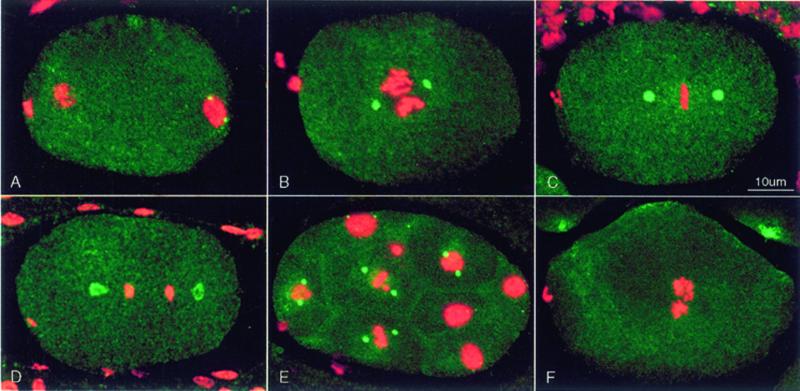
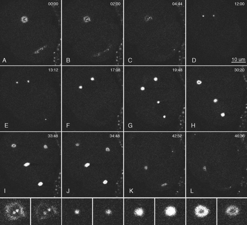
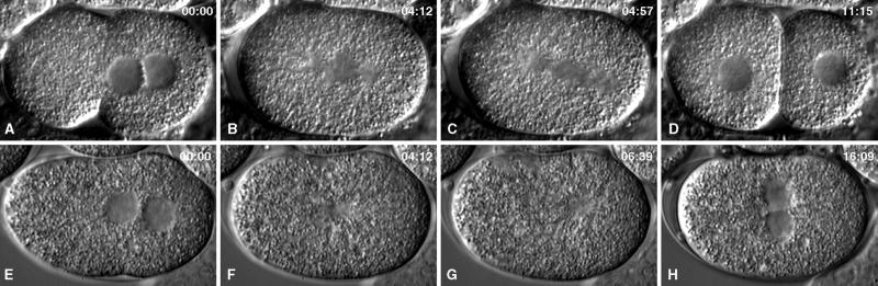
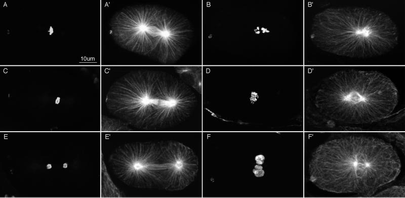
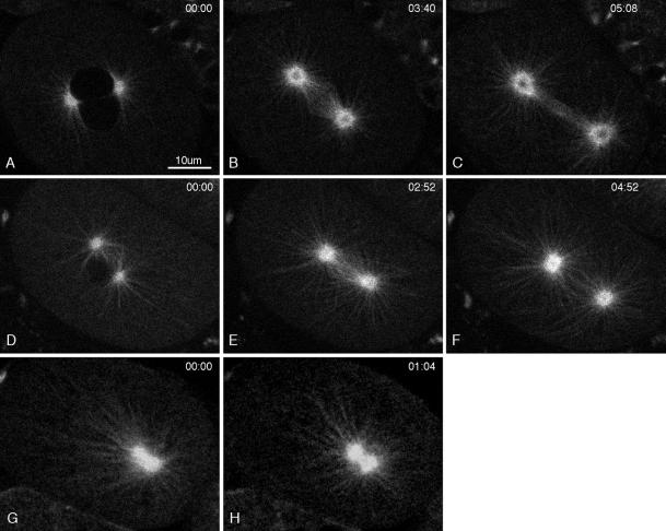
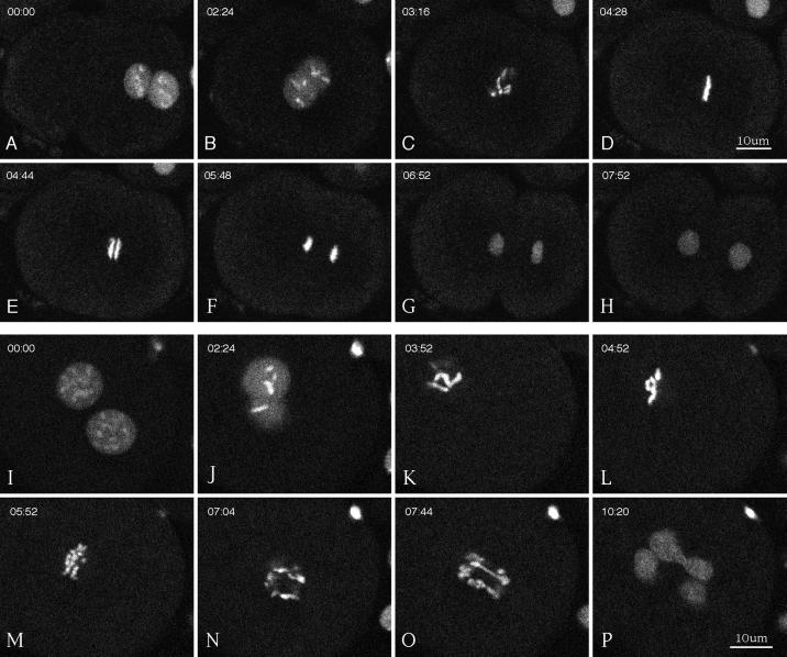
gamma-Tubulin is a ubiquitous and highly conserved component of centrosomes in eukaryotic cells. Genetic and biochemical studies have demonstrated that gamma-tubulin functions as part of a complex to nucleate microtubule polymerization from centrosomes. We show that, as in other organisms, Caenorhabditis elegans gamma-tubulin is concentrated in centrosomes. To study centrosome dynamics in embryos, we generated transgenic worms that express GFP::gamma-tubulin or GFP::beta-tubulin in the maternal germ line and early embryos. Multiphoton microscopy of embryos produced by these worms revealed the time course of daughter centrosome appearance and growth and the differential behavior of centrosomes destined for germ line and somatic blastomeres. To study the role of gamma-tubulin in nucleation and organization of spindle microtubules, we used RNA interference (RNAi) to deplete C. elegans embryos of gamma-tubulin. gamma-Tubulin (RNAi) embryos failed in chromosome segregation, but surprisingly, they contained extensive microtubule arrays. Moderately affected embryos contained bipolar spindles with dense and long astral microtubule arrays but with poorly organized kinetochore and interpolar microtubules. Severely affected embryos contained collapsed spindles with numerous long astral microtubules. Our results suggest that gamma-tubulin is not absolutely required for microtubule nucleation in C. elegans but is required for the normal organization and function of kinetochore and interpolar microtubules.


Figures

Figure 1
Figure 1
pie-1::GFP expression vector. Noncoding sequences from pie-1, a maternally expressed gene, were used to construct a vector that will express GFP fusion proteins in the adult germ line and in early embryos. A unique SpeI site was used to fuse open reading frames of interest to the carboxyl terminus of GFP.
Figure 2
Figure 2
Alignment of γ-tubulin sequences. (A) Sequences are from Homo sapiens (HS; GenBank accession number AF225971), Xenopus laevis (XL; M63446), Drosophila melanogaster (DM; AJ010552), Aspergillus nidulans (AN; X15479), and C. elegans (CE, CAA80164.1). Amino acids on black backgrounds are shared by three or more of the γ-tubulins shown. The open boxes enclose the C. elegans peptide sequences used to generate antibodies. (B) Grid showing the percent amino acid identities between pairs of γ-tubulins.
Figure 3
Figure 3
Distribution of γ-tubulin seen by immunofluorescence. Fixed embryos were stained with affinity-purified rabbit anti-γ-tubulin (green) and monoclonal PA3 anti-nucleosome antibody (red), which reveals chromatin. Each image is a projection of a Z series from a scanning confocal fluorescence microscope. Embryos in this and all subsequent figures are oriented with anterior to the left. (A) Early pronuclear migration. γ-Tubulin staining is concentrated in 2 small centrosomes associated with the male pronucleus at the posterior end. Centrosomes are not seen associated with the female pronucleus or polar body at the anterior end. (B) The pronuclei have met, and a prometaphase spindle has started rotation to align on the anterior–posterior axis. Centrosomes have recruited more γ-tubulin and appear larger. (C) In metaphase, the chromosomes are tightly packed on the spindle equator, and centrosomes have enlarged. (D) In anaphase, the centrosomal γ-tubulin has taken the form of hollow spheroids. The posterior centrosome is beginning to flatten. (E) Multicellular embryo showing pairs of bright γ-tubulin–containing centrosomes in mitotic cells. Interphase cells contain tiny γ-tubulin–containing centrosomes (e.g., top surface of the uppermost nucleus). (F) Detectable anti-γ-tubulin staining of centrosomes was eliminated by RNAi using dsRNA corresponding to the C. elegans γ-tubulin sequence.
Figure 4
Figure 4
Multiphoton images of GFP::γ-tubulin in a live wild-type embryo. Anterior is top left. Images were collected in a single focal plane (∼0.5 μm thick), and the elapsed time from the first frame is shown in minutes:seconds. (A and B) One-cell embryo during telophase. The anterior centrosome was a hollow sphere (a ring in cross section), and the posterior centrosome was flattening. Within each ring, 2 small dots of GFP::γ-tubulin were seen that grew into the centrosomes of the next cell cycle. (C) The flattened posterior centrosome dispersed before the anterior centrosome. In this frame and others, some daughter centrosomes are not visible, because they moved out of the focal plane. (D–F) In the anterior daughter cell (AB), the new centrosomes enlarged and migrated to opposite sides of the nucleus. (G) In the posterior daughter cell (P1), the centrosomes enlarged, migrated to opposite sides of the nucleus, and underwent the typical 90-deg rotation to align on the anterior–posterior axis. Mitosis in the anterior AB cell preceded that in P1. (H–L) Although more difficult to see in the second round of mitosis, the GFP::γ-tubulin signal was lost from the middle of each centrosome, and new daughter centrosomes appeared within the dark centers. The posterior centrosome in P1 repeated the pattern of flattening and early disappearance observed during the division of P0 (K and L). Bottom row, enlarged views of the top left centrosome from the period covered in B–I. Note the dispersal of the “mother” centrosome, appearance of daughter centrosomes within the mother ring, increase in centrosome size during mitosis, and reappearance of a dark center during the second anaphase.
Figure 5
Figure 5
Multiphoton image series of GFP::β-tubulin in a live wild-type embryo. Elapsed time from the first frame is shown in minutes:seconds. Brightness and contrast were adjusted to better reveal single microtubules in the top row and centrosome dynamics in the bottom 3 rows. Microtubule dynamics can be seen best in Movies 2 and 3. (A–C) Metaphase and early anaphase. (D–L) Anaphase B and telophase. The posterior spindle pole underwent dramatic side-to-side swinging during spindle elongation (C–G). Many astral microtubules extended to near the cortex. They usually appeared “swept back” on the side from which the spindle pole was moving (D). Some microtubules bent on the side to which the spindle pole was moving (see Movies 2 and 3). Note the bright area at the equator (D–F), which likely represents the region of interdigitation of interpolar microtubules. (G–L) Telophase and cytokinesis. The dark centers of the centrosomes enlarged, and the posterior mother centrosome flattened and fragmented. Two daughter centrosomes appeared within each dark center.
Figure 6
Figure 6
Comparison of a wild-type embryo (A–D) and a severely affected γ-tubulin(RNAi) embryo (E–H) during the first cell cycle. In these Nomarski images, nuclei (A, D, E, and H) and spindles (B, C, F, and G) are seen as granule-free zones. The top row shows a wild-type embryo at pronuclear meeting (A), prometaphase–metaphase (B), anaphase (B), and after cytokinesis (D). The bottom row shows corresponding stages in a γ-tubulin(RNAi) embryo. RNAi prevented the formation of a bipolar spindle, prevented normal chromosome segregation, and prevented cytokinesis.
Figure 7
Figure 7
Anti-tubulin and anti-histone immunofluorescence images of wild-type embryos (left 2 columns) and γ-tubulin(RNAi) embryos (right 2 columns). Fixed embryos were stained with rabbit anti-acetylated histone H4 antibodies to reveal chromatin (A–F) and mouse anti-α-tubulin antibodies to reveal microtubules (A′–F′). Each image is a projection of 5–7 sequential optical sections collected with a scanning confocal fluorescence microscope. Wild-type embryos are shown in prometaphase (A and A′), early anaphase (C and C′), and telophase (E and E′). (B and B′) Severely affected γ-tubulin(RNAi) embryo. Numerous long astral microtubules emanate from a collapsed spindle; chromosomes were not organized. (D and D′) Less severely affected RNAi embryo with a partially collapsed and poorly organized central spindle. Chromosomes appeared to be in early anaphase and were not well organized. (F and F′) RNAi embryo at telophase. The spindle had not segregated the chromosomes and showed little sign of interpolar microtubule bundles or spindle elongation. Chromatin was decondensing in micronuclei at the center of the embryo.
Figure 8
Figure 8
Multiphoton movies of GFP:: β-tubulin in a wild-type embryo (A–C), in a moderate γ-tubulin(RNAi) embryo (D–F), and in a severe γ-tubulin(RNAi) embryo (G–H). Elapsed time from the first frame is shown in minutes:seconds. (A and D) Prophase. The stage in D was further advanced than in A, and the 2 pronuclei were stacked one above the other. (B, E, and G) Metaphase, although the precise stage in the RNAi embryos (E and G) is difficult to determine. Both E and G were immediately before the spindle gyrations seen in RNAi embryos that we interpret as anaphase. (C and F) Anaphase B spindle elongation. Notice that an ordered bundle of interpolar microtubules is not seen in the moderate RNAi embryo (F). (H) In the severe embryo, the poles of the collapsed spindle did not separate. Comparison of G and H shows that the microtubule pattern changed with time, suggesting that even in severe γ-tubulin(RNAi) embryos, centrosomal microtubules are dynamic (also see Movie 4).
Figure 9
Figure 9
Multiphoton movies of GFP::histone in a wild-type embryo (A–H) and in a moderate γ-tubulin(RNAi) embryo (I–P). Elapsed time from the first frame is shown in minutes:seconds. (A, B, I, and J) Meeting, centering, and rotation of the pronuclei and prophase chromosome condensation. (C and K) Breakdown of the nuclear envelopes. (D and L) Metaphase. Tight alignment of chromosomes is shown on the metaphase plate in a wild-type embryo (D) versus loose alignment in the γ-tubulin(RNAi) embryo (L). (E and M) Early anaphase. (F and N) Late anaphase. (G, H, O, and P) Telophase. Note the poor segregation of chromosomes in the γ-tubulin(RNAi) embryo (N–P). Cytokinesis occurred in the wild-type embryo (H) but not in the RNAi embryo (P).

References

    1. Albertson DG. Formation of the first cleavage spindle in nematode embryos. Dev Biol. 1984;101:61–72. - PubMed
    1. Albertson DG, Thomson JN. Segregation of holocentric chromosomes at meiosis in the nematode Caenorhabditis elegans. Chromosome Res. 1993;1:15–26. - PubMed
    1. Bobinnec Y, Fukuda M, Nishida E. Identification and characterization of Caenorhabditis elegansgamma-tubulin in dividing cells and differentiated tissues. J Cell Sci. 2000;113:3747–3759. - PubMed
    1. Brenner S. The genetics of Caenorhabditis elegans. Genetics. 1974;77:71–94. - PMC - PubMed
    1. Consortium, T.C.e.S. Genome sequence of the nematode C. elegans: a platform for investigating biology. Science. 1998;282:2012–2018. - PubMed

Publication types

MeSH terms

LinkOut - more resources