. 2001 Jun;12(6):1843-57.
doi: 10.1091/mbc.12.6.1843.

Myosin vb is associated with plasma membrane recycling systems

Affiliations
Free PMC article

Myosin vb is associated with plasma membrane recycling systems

L A Lapierre et al. Mol Biol Cell. 2001 Jun.
Free PMC article

Abstract

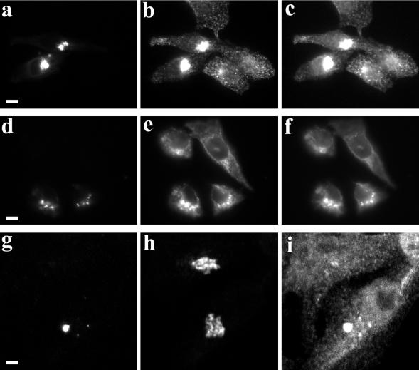
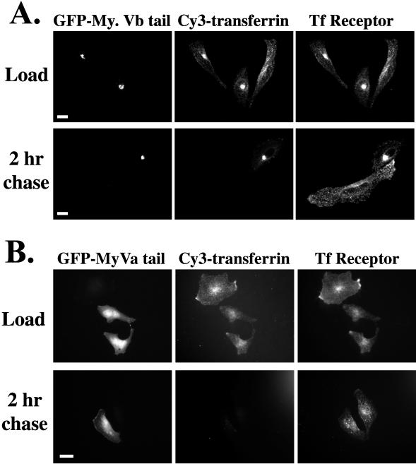
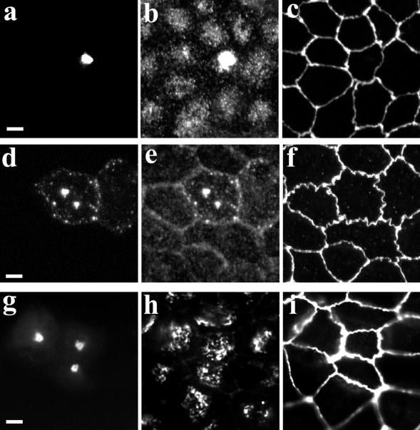
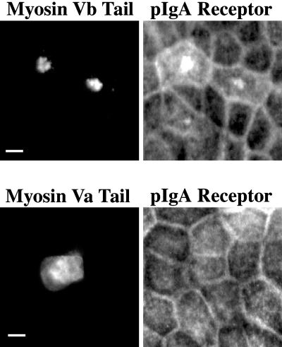
Myosin Va is associated with discrete vesicle populations in a number of cell types, but little is known of the function of myosin Vb. Yeast two-hybrid screening of a rabbit parietal cell cDNA library with dominant active Rab11a (Rab11aS20V) identified myosin Vb as an interacting protein for Rab11a, a marker for plasma membrane recycling systems. The isolated clone, corresponding to the carboxyl terminal 60 kDa of the myosin Vb tail, interacted with all members of the Rab11 family (Rab11a, Rab11b, and Rab25). GFP-myosin Vb and endogenous myosin Vb immunoreactivity codistributed with Rab11a in HeLa and Madin-Darby canine kidney (MDCK) cells. As with Rab11a in MDCK cells, the myosin Vb immunoreactivity was dispersed with nocodazole treatment and relocated to the apical corners of cells with taxol treatment. A green fluorescent protein (GFP)-myosin Vb tail chimera overexpressed in HeLa cells retarded transferrin recycling and caused accumulation of transferrin and the transferrin receptor in pericentrosomal vesicles. Expression of the myosin Vb tail chimera in polarized MDCK cells stably expressing the polymeric IgA receptor caused accumulation of basolaterally endocytosed polymeric IgA and the polymeric IgA receptor in the pericentrosomal region. The myosin Vb tail had no effects on transferrin trafficking in polarized MDCK cells. The GFP-myosin Va tail did not colocalize with Rab11a and had no effects on recycling system vesicle distribution in either HeLa or MDCK cells. The results indicate myosin Vb is associated with the plasma membrane recycling system in nonpolarized cells and the apical recycling system in polarized cells. The dominant negative effects of the myosin Vb tail chimera indicate that this unconventional myosin is required for transit out of plasma membrane recycling systems.


Figures

Figure 1
Figure 1
Partial amino acid sequence of rabbit myosin Vb compared with rat myosin Vb (myr 6), a murine myosin Vb fragment mistakenly identified as glutamate decarboxylase (GAD) and chicken myosin Va. Underlined regions denote amino acid sequences required for association with Rab11a (Figure 2).
Figure 2
Figure 2
Rab11a interaction with truncated myosin Vb tail constructs. The structure of the myosin Vb tail is shown schematically including a region of coiled-coil structure and the terminal globular region. Amino acid numbers in parentheses correspond to the sequence of murine myosin Vb (myr 6), whereas the nonbracketed numbers refer to the amino acid sequence of the rabbit myosin Vb tail clone shown in Figure 1. Truncations of the myosin Vb tail sequence were assembled in pADGAL and cotransfected into yeast with either pBDGAL-Rab11a or pBDGAL-Rab11aS20V. Lifts of yeast colonies were then exposed to Xgal and development of blue color was interpreted as a positive interaction (+). Results with Rab11aS20V were identical to those with Rab11a wild type.
Figure 3
Figure 3
Localization of GFP-myosin Vb in transfected HeLa and MDCK cells. HeLa cells (a–c) were transiently transfected with a full-length GFP-myosin Vb chimera. Cells were triple imaged for GFP-myosin Vb (a), Rab11a (b), and transferrin receptor (c). Arrowheads indicate the positions of cell-labeled extensions in transfected cells. MDCK cells (d–f) were transfected with the full-length GFP-myosin Vb chimera and grown at confluence for 4 d on permeable filters. Cells were triple imaged for GFP-myosin Vb (d), Rab11a (e), and ZO-1 (f). Images in MDCK cells represent projections assembled from confocal optical section series. The results are representative of three separate experiments. Bar, 5 μm (a–c), 1 μm (d–f).
Figure 4
Figure 4
Localization of endogenous myosin Vb in MDCK cells stably expressing GFP-Rab11a. GFP-Rab11a/MDCK cells were grown at confluence for 4 d on permeable filters. Cells were fixed after no treatment (a–c), treatment with nocodazole (d–f), or treatment with taxol (g–i). Cells were triple imaged for GFP-Rab11a (a, d, and g), endogenous myosin Vb immunostaining (b, e, and h), and ZO-1 immunostaining (c, f, and i). Each panel represents X-Y confocal projection reconstructions. Arrowheads indicate the position used for X-Z reconstructions shown in the bottom portion of each panel. The results are representative of two separate experiments. Bar, 1 μm (a–c), 2 μm (d–i).
Figure 5
Figure 5
Morphological effects of overexpression of GFP-myosin Vb tail in HeLa cells. (a–f) HeLa cells were transiently transfected with GFP-myosin Vb tail and allowed to recover for 24 h. Cells then received either no treatment (a–c) or treatment with nocodazole (d–f). Cells were then fixed and in addition to imaging GFP (a and d) cells were stained for endogenous transferrin receptor (b and e) and endogenous Rab11a (c and f). (g and h) HeLa cells were transiently transfected with GFP-myosin Vb tail as described above. Fixed cells were triple imaged for GFP (g), p58 immunostaining, a Golgi marker (h), and Rab11a immunostaining (i). Images in g–i represent projections assembled from fifteen 0.29-μm optical sections obtained in confocal microscopy. The results are representative of three separate experiments. Bar, 5 μm (a–f), 2 μm (g–i).
Figure 6
Figure 6
Effects of overexpression of GFP-myosin Vb tail and GFP-myosin Va tail on transferrin trafficking in HeLa cells. To assess the effects on transferrin trafficking GFP-myosin Vb tail (A) or GFP-myosin Va tail (B) were transiently transfected into HeLa cells. Twenty-four hours after transfection, cells were serum starved for 30 min and then allowed to endocytose Cy3-transferrin for 60 min. After uptake, some cells were allowed to recover in 10% fetal bovine serum-containing medium for 2 h. Cells were imaged for GFP, Cy3-transferrin, and immunostaining for endogenous transferrin receptor. The results are representative of four separate experiments. Bar, 5 μm.
Figure 7
Figure 7
Effects of GFP-myosin Vb on recycling of biotinylated transferrin in HeLa cells. Nontransfected HeLa cells (NT) or HeLa cells transfected with either GFP-myosin Va tail (Va) or GFP-myosin Vb tail (Vb) were loaded with biotinylated-human transferrin. After washing, cells were allowed to recover for either 5 or 20 min in media containing unlabeled transferrin. Electrophoretic transfers of cell lysates were incubated with horseradish peroxidase-streptavidin to visualize transferrin (A) Triplicate samples are shown. (B) Results were quantified and expressed as a percentage of transferrin retained within cells after 20 min (mean + SEM; n = 6). *p < 0.01 compared with both NT and myosin Va by Mann-Whitney test.
Figure 8
Figure 8
GFP-myosin Vb tail alters apical recycling system morphology in MDCK cells. MDCK were transiently transfected with GFP-myosin Vb tail and grown for 3 d at confluence on permeable filters. (a–f) Cells were then exposed to either no treatment (a–c) or treatment with nocodazole (d–f). Fixed cells were triple imaged for GFP (a and d), endogenous Rab11a (b and e), and ZO-1 (c and f). (g–i) GFP-myosin Vb tail transfected MDCK cells were fixed and triple imaged for GFP (g), the Golgi marker p58 (h), and ZO-1 (i). The results are representative of three separate experiments. Bar, 5 μm.
Figure 9
Figure 9
GFP-myosin Vb tails alters the distribution of polymeric IgA receptor. MDCK cells were transiently transfected with either GFP-myosin Vb tail or GFP-myosin Va tail and grown for 3 d at confluence on permeable filters. Cells were then fixed and dual imaged for GFP and pIgR immunostaining. The results are representative of three separate experiments. Bar, 5 μm.
Figure 10
Figure 10
Alteration of trafficking in MDCK cells by GFP-myosin Vb tail. (a–f) MDCK cells stably expressing the polymeric IgA receptor were transiently transfected with GFP-myosin Vb tail and allowed to grow at confluence on permeable filters for 4 d. The cells were then loaded with Texas Red-IgA from the basolateral media for 60 min and then either fixed (a–c) or allowed to recover in fresh medium as a chase for 60 min (d–f). Cells were triple imaged for GFP (a and d), Texas Red-IgA (b and e), and endogenous Rab11a immunostaining (c and f). (g–i) MDCK cells were transiently transfected with GFP-myosin Vb tail and allowed to grow at confluence on permeable filters for 4 d. Cells were loaded with Cy3-transferrin for 60 min and fixed. Cells were triple imaged for GFP (g), Cy3-transferrin (h), and endogenous Rab11a immunostaining (i). All panels represent X-Y reconstructions of confocal optical section series. Arrowhead marks the location used for assembly of X-Z projections seen below X-Y projections in g–i. Bar, 5 μm (a–f), 2 μm (g–i).

References

    1. Apodaca G, Katz LA, Mostov KE. Receptor-mediated transcytosis of IgA in MDCK cells is via apical recycling endosomes. J Cell Biol. 1994;125:67–86. - PMC - PubMed
    1. Beningo KA, Lillie SH, Brown SS. The yeast kinesin-related protein Smy1p exerts its effects on class V myosin Myo2p via a physical interaction. Mol Biol Cell. 2000;11:691–702. - PMC - PubMed
    1. Bhartur SG, Goldenring JR. Mapping of ezrin dimerization using yeast two-hybrid screening. Biochem Biophys Res Commun. 1998;243:874–877. - PubMed
    1. Bonafe N, Sellers JR. Molecular characterization of myosin V from Drosophila melanogaster. J Muscle Res Cell Motil. 1998;19:129–141. - PubMed
    1. Bridgman PC. Myosin Va movements in normal and dilute-lethal axons provide support for a dual filament motor complex. J Cell Biol. 1999;146:1045–1060. - PMC - PubMed

Publication types

MeSH terms