. 2001:2:4.
doi: 10.1186/1471-2172-2-4. Epub 2001 Jun 8.

A conditional form of Bruton's tyrosine kinase is sufficient to activate multiple downstream signaling pathways via PLC Gamma 2 in B cells

Affiliations

A conditional form of Bruton's tyrosine kinase is sufficient to activate multiple downstream signaling pathways via PLC Gamma 2 in B cells

M G Tomlinson et al. BMC Immunol. 2001.

Abstract

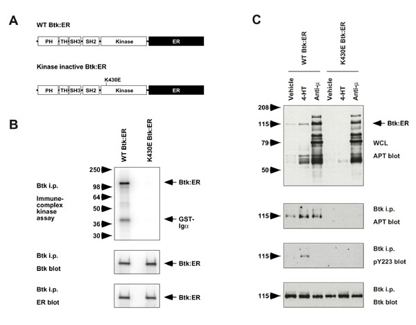
Background: Bruton's tyrosine kinase (Btk) is essential for B cell development and function. Mutations of Btk elicit X-linked agammaglobulinemia in humans and X-linked immunodeficiency in the mouse. Btk has been proposed to participate in B cell antigen receptor-induced signaling events leading to activation of phospholipase C-gamma2 (PLCgamma2) and calcium mobilization. However it is unclear whether Btk activation is alone sufficient for these signaling events, and whether Btk can activate additional pathways that do not involve PLCgamma2. To address such issues we have generated Btk:ER, a conditionally active form of the kinase, and expressed it in the PLCgamma2-deficient DT40 B cell line.

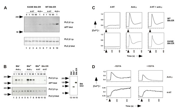
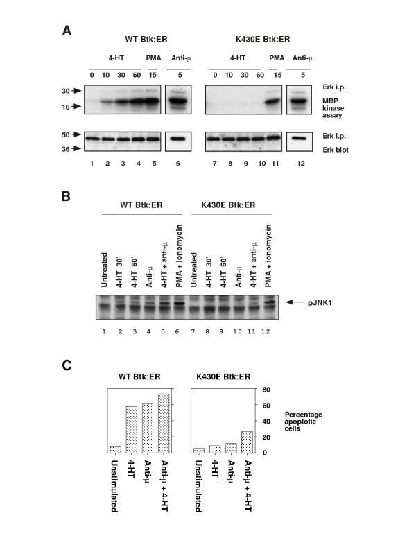
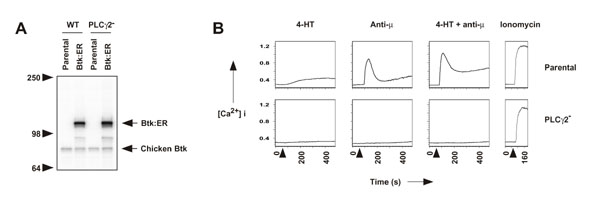
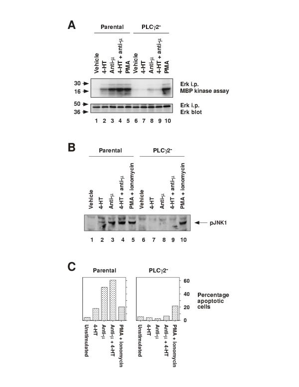
Results: Activation of Btk:ER was sufficient to induce multiple B cell signaling pathways in PLCgamma2-sufficient DT40 cells. These included tyrosine phosphorylation of PLCgamma2, mobilization of intracellular calcium, activation of extracellular signal-regulated kinase (ERK) and c-Jun NH2-terminal kinase (JNK) mitogen-activated protein kinase (MAPK) pathways, and apoptosis. In DT40 B cells deficient for PLCgamma2, Btk:ER activation failed to induce the signaling events described above with the consequence that the cells failed to undergo apoptosis.

Conclusions: These data suggest that Btk:ER regulates downstream signaling pathways primarily via PLCgamma2 in B cells. While it is not known whether activated Btk:ER precisely mimics activated Btk, this conditional system will likely facilitate the dissection of the role of Btk and its family members in a variety of biological processes in many different cell types.


Figures

Figure 1
Figure 1
Expression of Btk:ER, a conditional form of Btk, in Btk-deficient DT40 cells. A, Diagrammatic representation of Btk:ER constructs. WT and kinase inactive (K430E) forms of Btk were fused at their COOH-terminus to the hormone-binding domain of the estrogen receptor (ER). B, Immune-complex kinase assay and Western blot analyses of Btk:ER constructs. Btk-deficient DT40 cells were stably transfected with WT Btk:ER or K430E Btk:ER and clones selected that expressed equal levels of Btk:ER. Cells were lysed and Btk:ER was immunoprecipitated with specific Btk antiserum. Immune complex kinase assays were performed using GST-Igα as an exogenous substrate (top panel), in addition to immunoblotting with antisera specific for Btk (middle panel) and ER (lower panel). C, Activation of Btk:ER in vivo by 4-HT. Cells expressing either WT or kinase inactive Btk:ER were stimulated with vehicle control, 4-HT (1 μM for 30-min) or anti-μmAb M4 (4 μg/ml for 1-min). Cells were lysed and Btk:ER was immunoprecipitated with specific Btk antiserum. Whole cell lysates were blotted with mAb 4G10 (top panel). Btk mmunoprecipitates were blotted with mAb 4G10, phosphospecific Btk Y223 antiserum, or Btk antiserum (lower panels).
Figure 2
Figure 2
Btk:ER activation is sufficient to induce PLCγ2 phosphorylation and calcium mobilization in Btk-deficient DT40 cells. A, Btk:ER-induced PLCγ2 phosphorylation. Cells expressing either WT or kinase inactive Btk:ER were lysed as a function of the indicated times (minutes) after addition of 4-HT (1 μM) or BCR cross-linking with anti-μ mAb M4 (4 μg/ml). PLCγ2 was immunoprecipitated with anti-PLCγ2 antiserum and immunoblotted with anti-phosphotyrosine mAb (APT, top panel) and with anti-PLCγ2 antiserum (bottom panel). B, BCR-induced PLCγ2 phosphorylation in Btk-deficient versus Btk-sufficient cells. The experiment was performed as described for Fig. 2A. Lanes 1-8 show anti-μ-induced PLCγ2 phosphorylation in Btk-deficient DT40 cells and in Btk-deficent cells expressing mouse Btk, which were described previously [13]. Lanes 9-12 show 4-HT-induced PLCγ2 phosphorylation in Btk-deficient cells expressing Btk or Btk:ER. Lanes 13-15 show a Btk blot of whole cell lysates for Btk-deficient cells, Btk-deficient cells expressing Btk, and Btk-deficient cells expressing Btk:ER. C, Btk:ER-induced calcium mobilization. Intracellular free calcium levels in indo-1-loaded cells were monitored over an 8-min period by FACS. 4-HT (1 μM) and/or anti-μ mAb M4 (2 μg/ml) were added at the 60 s time point. Relative intracellular calcium levels are shown on the y-axis. D, Sustained calcium mobilization is dependent on calcium influx. Intracellular calcium levels were monitored as described in panel B, in the presence or absence of 3 mM EGTA which chelates extracellular calcium. These data are representative of three independent stable clones.
Figure 3
Figure 3
Btk:ER activation is sufficient to induce ERK and JNK activation, and apoptosis, in Btk-deficient DT40 cells. A, Btk:ER-induced ERK MAPK activation. Cells were lysed as a function of the indicated times (minutes) after addition of 4-HT (1 μM), anti-μ mAb M4 (4 μg/ml), or phorbol 12-myristate 13-acetate (PMA, 100 ng/ml) as a positive control. ERK1 and ERK2 were immunoprecipitated with ERK1 and ERK2 antisera. Immune complex kinase assays were performed using myelin basic protein as an exogenous substrate (top panel). Samples were immunoblotted with anti-ERK1 and ERK2 antisera to show equal loading (bottom panel). ERK1 and ERK2 were not resolved as individual bands in these experiments, and ERK2 was the major species detected (data not shown). B, Btk:ER-induced JNK phosphorylation. Cells were lysed as a function of the indicated times (minutes) after addition of 4-HT (1 μM) and/or anti-μ mAb M4 (4 μg/ml), or PMA (100 ng/ml) and ionomycin (250 ng/ml) as a positive control. Whole cell lysates were immunoblotted with anti-active JNK phosphospecific antiserum. C, Btk:ER-induced apoptosis. Cells expressing either WT or kinase inactive Btk:ER were cultured for 24 h with 4-HT (1 μM) and/or anti-μ mAb M4 (2 μg/ml). Apoptosis was measured by TUNEL assay. The percentage of TUNEL-positive cells was calculated with reference to a negative control TUNEL reaction in the absence of terminal transferase. Negative controls yielded less than 1% TUNEL-positive cells in all cases. These data are representative of at least three independent stable clones.
Figure 4
Figure 4
Btk:ER-induced calcium mobilization is dependent on PLCγ2. A, expression of Btk:ER in parental and PLCγ2-deficient DT40 cells. WT Btk:ER was transfected into parental and PLCγ2-deficient cells and stable clones selected that expressed equal levels of Btk:ER. Whole cell lysates were immunoblotted with antiserum specific for Btk. B, Btk:ER-induced calcium mobilization is dependent on PLCγ2. Calcium mobilization in parental and PLCγ2-deficient cells expressing Btk:ER was measured as described in the legend for Fig 2C. Ionomycin (250 ng/ml) was used as a positive control.
Figure 5
Figure 5
Btk:ER-induced ERK and JNK activation, and apoptosis, are dependent on PLCγ2. A, Btk:ER-induced ERK MAPK activation is dependent on PLCγ2. Parental and PLCγ2-deficient cells expressing Btk:ER were treated for 60-min with vehicle control (0.1% ethanol), 4-HT (1 μM) and/or anti-μ mAb M4 (4 μg/ml), or for 15-min with PMA (100 ng/ml) as a positive control. ERK activation was measured by immune complex kinase assay as described in the legend for Fig 3A. B, Btk:ER-induced JNK phosphorylation is dependent on PLCγ2. Parental and PLCγ2-deficient cells expressing Btk:ER were treated for 30-min with vehicle control (0.1% ethanol), 4-HT (1 μM) and/or anti-μ mAb M4 (4 μg/ml), or for 10-min with PMA (100 ng/ml) and ionomycin (250 ng/ml) as a positive control. JNK phosphorylation was measured by immunblotting with anti-active JNK phosphospecific antiserum as described in the legend for Fig 3B. C, Btk:ER-induced apoptosis is dependent on PLCγ2. Apoptosis of parental and PLCγ2-deficient cells expressing Btk:ER was measured by TUNEL assay as described in the legend for Fig 3C. PMA (100 ng/ml) and ionomycin (250 ng/ml) were used as a positive control. These data are representative of three independent stable clones.

Similar articles

Cited by

References

    1. Satterthwaite AB, Witte ON. The role of Bruton's tyrosine kinase in B-cell development and function: a genetic perspective. Immunol Rev. 2000;175:120–127. doi: 10.1034/j.1600-065X.2000.017504.x. - DOI - PubMed
    1. Schaeffer EM, Schwartzberg PL. Tec family kinases in lymphocyte signaling and function. Curr Opin Immunol. 2000;12:282–288. doi: 10.1016/S0952-7915(00)00088-1. - DOI - PubMed
    1. Yang WC, Collette Y, Nunes JA, Olive D. Tec kinases: a family with multiple roles in immunity. Immunity. 2000;12:373–382. - PubMed
    1. Kawakami Y, Kitaura J, Hata D, Yao L, Kawakami T. Functions of Bruton's tyrosine kinase in mast and B cells. J Leukoc Biol. 1999;65:286–290. - PubMed
    1. Mohamed AJ, Nore BF, Christensson B, Smith CI. Signalling of Bruton's tyrosine kinase, Btk. Scand J Immunol. 1999;49:113–118. doi: 10.1046/j.1365-3083.1999.00504.x. - DOI - PubMed

Publication types