. 2001 Jun 15;15(12):1493-505.
doi: 10.1101/gad.892301.

casanova encodes a novel Sox-related protein necessary and sufficient for early endoderm formation in zebrafish

Affiliations

casanova encodes a novel Sox-related protein necessary and sufficient for early endoderm formation in zebrafish

Y Kikuchi et al. Genes Dev. .

Abstract

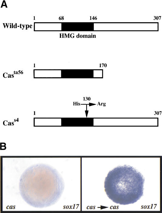
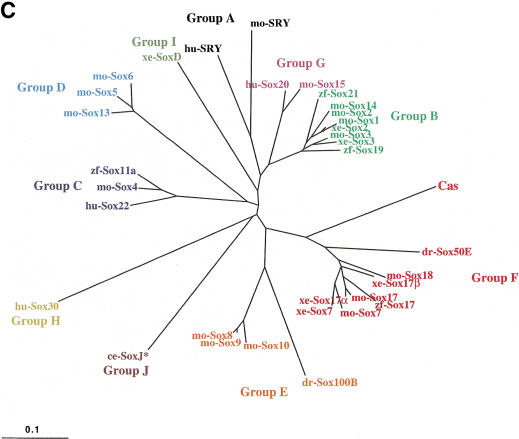
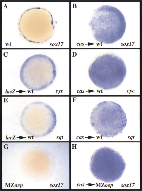
Early endoderm formation in zebrafish requires at least three loci that function downstream of Nodal signaling but upstream of the early endodermal marker sox17: bonnie and clyde (bon), faust (fau), and casanova (cas). cas mutants show the most severe phenotype as they do not form any gut tissue and lack all sox17 expression. Activation of the Nodal signaling pathway or overexpression of Bon or Fau/Gata5 fails to restore any sox17 expression in cas mutants, demonstrating that cas plays a central role in endoderm formation. Here we show that cas encodes a novel member of the Sox family of transcription factors. Initial cas expression appears in the dorsal yolk syncytial layer (YSL) in the early blastula, and is independent of Nodal signaling. In contrast, endodermal expression of cas, which begins in the late blastula, is regulated by Nodal signaling. Cas is a potent inducer of sox17 expression in wild-type embryos as well as in bon and fau/gata5 mutants. Cas is also a potent inducer of sox17 expression in MZoep mutants, which cannot respond to Nodal signaling. In addition, ectopic expression of cas in presumptive mesodermal cells leads to their transfating into endoderm. Altogether, these data indicate that Cas is the principal transcriptional effector of Nodal signaling during zebrafish endoderm formation.


Figures

Figure 1
Figure 1
cas encodes a novel member of the Sox family of high-mobility group (HMG) domain transcription factors. (A) Schematic representation of wild-type and mutant Cas proteins. The casta56 mutation leads to a truncation of the protein shortly after the HMG domain, and the cass4 mutation introduces a single amino acid substitution (histidine to arginine) in the third helix of the HMG domain (position 130). (B) sox17 expression in uninjected (left) and CG569/cas-injected casta56mutant embryos. Animal pole views at shield stage (6 h postfertilization) with dorsal to the right (left embryo). Whereas sox17 expression is missing in uninjected casta56mutants (left), overexpression of CG569/cas mRNA always induced sox17 expression (right). For comparison to wild-type pattern of sox17 expression, see Fig. 4A. (C) Unrooted phylogenetic tree for the Sox HMG domain, constructed by the neighbor joining (NJ) method (Saitou and Nei 1987). Branch lengths are representative of the extent of divergence. Ten groups (Groups A–J) are classified by phylogenetic analysis of Sox protein HMG domain sequences (Bowles et al. 2000). The ce-SoxJ* protein (Group J) was identified from databases and previously reported as a Sox protein (Bowles et al. 2000). This phylogenetic analysis indicates that Cas is a divergent member of the SoxF group. (D) Sequence comparison of Cas; zebrafish, Xenopus and mouse Sox17; mouse Sox18; and Xenopus and mouse Sox7. The HMG domain is underlined. Cas shows a relatively high degree of sequence identity in its HMG domain with the members of the SoxF group, but otherwise shows limited sequence conservation. The arrow indicates intron position in cas, zebrafish sox17, and mouse Sox17 and Sox18, which appears to be the same in all members of the SoxF group but different in most other Sox genes (Bowles et al. 2000). The asterisk and exclamation point indicate the position of the cass4 and casta56 mutations, respectively. (E) Percent sequence identity of the Cas and zebrafish Sox17 HMG domains with the HMG domains of the other members of the SoxF group as well as with the HMG domains of mouse Sox10 (SoxE group) and mouse LEF1. Cas shows substantially less sequence identity with the other SoxF group members than does zebrafish Sox17. However, Cas shows even less identity with mouse Sox10 (54%) and mouse LEF1 (29%). Abbreviations: ce, Caenorhabditis elegans; dr, Drosophila melanogaster; hu, Homo sapiens; mo, mouse; xe, Xenopus laevis; zf, Danio rerio.
Figure 1
Figure 1
cas encodes a novel member of the Sox family of high-mobility group (HMG) domain transcription factors. (A) Schematic representation of wild-type and mutant Cas proteins. The casta56 mutation leads to a truncation of the protein shortly after the HMG domain, and the cass4 mutation introduces a single amino acid substitution (histidine to arginine) in the third helix of the HMG domain (position 130). (B) sox17 expression in uninjected (left) and CG569/cas-injected casta56mutant embryos. Animal pole views at shield stage (6 h postfertilization) with dorsal to the right (left embryo). Whereas sox17 expression is missing in uninjected casta56mutants (left), overexpression of CG569/cas mRNA always induced sox17 expression (right). For comparison to wild-type pattern of sox17 expression, see Fig. 4A. (C) Unrooted phylogenetic tree for the Sox HMG domain, constructed by the neighbor joining (NJ) method (Saitou and Nei 1987). Branch lengths are representative of the extent of divergence. Ten groups (Groups A–J) are classified by phylogenetic analysis of Sox protein HMG domain sequences (Bowles et al. 2000). The ce-SoxJ* protein (Group J) was identified from databases and previously reported as a Sox protein (Bowles et al. 2000). This phylogenetic analysis indicates that Cas is a divergent member of the SoxF group. (D) Sequence comparison of Cas; zebrafish, Xenopus and mouse Sox17; mouse Sox18; and Xenopus and mouse Sox7. The HMG domain is underlined. Cas shows a relatively high degree of sequence identity in its HMG domain with the members of the SoxF group, but otherwise shows limited sequence conservation. The arrow indicates intron position in cas, zebrafish sox17, and mouse Sox17 and Sox18, which appears to be the same in all members of the SoxF group but different in most other Sox genes (Bowles et al. 2000). The asterisk and exclamation point indicate the position of the cass4 and casta56 mutations, respectively. (E) Percent sequence identity of the Cas and zebrafish Sox17 HMG domains with the HMG domains of the other members of the SoxF group as well as with the HMG domains of mouse Sox10 (SoxE group) and mouse LEF1. Cas shows substantially less sequence identity with the other SoxF group members than does zebrafish Sox17. However, Cas shows even less identity with mouse Sox10 (54%) and mouse LEF1 (29%). Abbreviations: ce, Caenorhabditis elegans; dr, Drosophila melanogaster; hu, Homo sapiens; mo, mouse; xe, Xenopus laevis; zf, Danio rerio.
Figure 2
Figure 2
cas expression in zebrafish embryos at high (3.3 h postfertilization [hpf]; A,B), sphere (4 hpf; C,D), 30% epiboly (4.7 hpf; I), 40% epiboly (5 hpf; E,F,N,O,P), 50% epiboly (5.3 hpf; J), 60% epiboly (7 hpf; K,L), and 90% epiboly (9 hpf; G,H,Q) stages. sqt expression at 60% epiboly (7 hpf; M). A, C, E, F, G, L, M, N, P, and Q show lateral views with dorsal to the right. B, D, and O show animal pole views. H, I, J, and K show dorsal views with anterior to the top. (A,B) cas expression is first observed at the high stage in the dorsal YSL. (C,D) By sphere stage cas expression is detected throughout the marginal (yolk syncytial layer) YSL and in dorsal blastomeres. (E,F) At 40% epiboly cas is expressed in a subset of blastomeres all around the margin (see high magnification in F), and is maintained in the marginal YSL. (G,H) As endodermal cells gastrulate, they continue to express cas, and a group of noninvoluting cells, the forerunner cells (fo), also begins to express cas. At 90% epiboly, expression is seen in all endodermal and forerunner cells and resembles sox17 expression. (I) cas expression in presumptive forerunner cells. (J,K) By 60% epiboly, forerunners, which continue to express cas, form a compact group of cells that do not involute and stay at the vegetal tip of the axial mesoderm throughout gastrulation. (L,M) Forerunners show strong cas expression (L) as well as sqt (M) expression throughout gastrulation. (N,O) Injection of 100 pg of taram-a* mRNA into one cell at the two-cell stage led to a high level of cas expression in the injected half of the embryo. (P,Q) Injection of a low dose of antivin (atv) mRNA (25 pg) at the one-cell stage led to the disappearance of embryonic cas expression, whereas the YSL expression appeared unaffected. High magnification views at 40% epiboly are shown in P. The optical cross section in Q shows cas expression present in the YSL, but absent in the endoderm of atv injected embryos at 90% epiboly. ap, animal pole; d, dorsal; end, endoderm; fo, forerunner cells; v, ventral; wt, wild type; ysl, yolk syncytial layer.
Figure 3
Figure 3
cas expression in cas, bon, and fau/gata5 mutant embryos. Animal pole views with dorsal to the right at shield stage (6 h postfertilization [hpf]). (A–C), and dorsal views with anterior to the top at 80% epiboly (8.3 hpf; D–F). (A,D) In cas mutants, no cas expression is observed in the embryo proper, whereas cas appears to be up-regulated in the yolk syncytial layer (YSL). (B,E) In bon mutants, cas expression is observed in a few endodermal cells (arrows), the forerunner cells (arrowheads), and the YSL. (C,F) In fau/gata5 mutants, cas expression in the endodermal cells appears weaker (arrows), whereas cas expression in the forerunner cells (arrowheads) and the YSL appears normal.
Figure 4
Figure 4
cas overexpression induces cyc and sqt expression in wild-type embryos, and induces sox17 expression in MZoep mutants. (A,B,G,H) Animal pole views at shield stage (6 h postfertilization [hpf]) with dorsal to the right (A). (C–F) Animal pole views at 50% epiboly (5.3 hpf) with dorsal to the right (C,E). (A,B) A characteristic salt-and-pepper distribution of sox17-expressing endodermal cells is observed throughout the margin in uninjected wild-type embryos (A), whereas injection of cas mRNA at the 1–4-cell stage led to confluent patches of sox17 expression throughout the embryo (B). (C–F) Injection of cas mRNA at the 1–4-cell stage induced ectopic cyc and sqt expression in wild-type embryos (D,F). (G,H) Whereas no sox17 expression is observed in uninjected MZoep mutants (G), injection of cas mRNA at the 1–4-cell stage induced strong sox17 expression (H). wt, wild type.
Figure 5
Figure 5
cas overexpression induces ectopic sox17 expression in bon and fau/gata5 mutant embryos, whereas bon and fau/gata5 cannot induce ectopic cas expression in wild-type embryos. Animal pole views at 6 h postfertilization (hpf; A–C) and 40% epiboly stage (5 hpf; D–F). (A–C) Injection of cas mRNA at the 1–4-cell stage expanded sox17 expression to a comparable extent in wild-type, bon, and fau/gata5 mutant embryos. (D–F) Injection of bon or fau/gata5 mRNA in wild-type embryos was not able to induce ectopic cas expression away from the margin.
Figure 6
Figure 6
Misexpression of cas results in the transfating of presumptive mesodermal cells into endoderm. GFP fluorescence at shield stage (6 hours postfertilization [hpf]; A; animal pole view; T; lateral view), and at 24 hpf (C,L,V; lateral views). Lateral view with dorsal to the right of sox17 expression at shield stage (B,W). gsc expression at 50% epiboly (5.3 hpf; F,G; animal pole views, dorsal to the right). hgg1 expression at 80% epiboly (8.3 hpf; H,I; dorsal views, anterior to the top). shh expression at 24 hpf (J,K; lateral views). carbonic anhydrase expression at 24 hpf (M,N; lateral views). Fate map, as adapted from Thisse et al. (2000) of uninjected (O) and cas-injected embryos (P–R) at the onset of gastrulation (5.3 hpf). sqt expression at 30% epiboly (4.7 hpf; X) and 40% epiboly (5 hpf; Y,Z); lateral views, dorsal to the right. (A) Coinjection of 100 pg of cas mRNA and 100 pg of GFP mRNA into a marginal blastomere at the 16-cell stage gives rise to marginal clones of cells which were screened at the onset of gastrulation to determine their localization along the dorso–ventral axis. A dorsal clone is shown here. (B) Embryo with a labeled dorsal clone was examined for sox17 expression, which was strongly induced dorsally. (C) Embryo shown in A at 24 hpf. GFP-expressing cells accumulate in the entire endoderm, from the level of the pharyngeal arches anteriorly to the anal opening in the posterior trunk region. More caudally, GFP-expressing cells are observed underlying the notochord in the anterior part of the tail, and in the notochord and tail bud more posteriorly. (D) High-magnification view of the head of the embryo shown in C, revealing a cyclopic phenotype. (E) The same embryo exhibits U-shaped somites, indicative of a reduction in axial mesoderm. (F,G) gsc expression in a dorsally injected embryo (G) is severely reduced as compared to wild type (F). (H,I) Expression of hgg1, a prechordal plate marker, in a dorsally-injected embryo (I) is nearly abolished as compared to wild type (H). (J,K) shh expression in a dorsally-injected embryos (K) is severely reduced as compared to wild type (J). (L) Embryo with a labeled ventral clone at 24 hpf. GFP-expressing cells accumulate in the tail, which displays an abnormal shape characterized by a large, empty ventral blister instead of ventral mesenchyme. (M,N) The blood expression of carbonic anhydrase has almost completely disappeared in a ventrally-injected embryo, whereas the otic vesicle (ot) expression is unaffected. (O) Fate map of a wild-type embryo at the onset of gastrulation (di, diencephalon; ep, epidermis; hb, hindbrain; mb, midbrain; n, notochord; pp, prechordal plate; sp, spinal chord; te, telencephalon). (P) Misexpression of cas in a clone of cells located in the central part of the embryonic shield results in the transfating of the prechordal plate into endoderm, resulting in the loss of gsc (G) and hgg1 (I) expression. (Q) A larger dorsal clone affects both anterior and posterior axial mesoderm, resulting in embryos that lack both prechordal and notochordal mesoderm, as shown in K. (R) Ventral clones result in embryos that lack almost all blood cells but appear unaffected in the other mesodermal derivatives, as shown in N. (S,T) An embryo injected with cas and GFP mRNA in an animal pole blastomere at the 256-cell stage (S) is shown at shield stage in T. (U,V) At 24 hpf, embryos injected at the animal pole have a wild-type morphology (U) and the GFP-expressing cells do not populate the endoderm but are often found on top of the yolk (V). (W,X) Injection of cas at the animal pole induces sox17 (W) but not sqt (X) expression in the embryo proper. In such embryos, sqt expression is only induced in a few cells of the enveloping layer (evl cells; e), an extraembryonic structure. (Y,Z) In contrast, marginal cas injection induces strong sqt expression in embryonic cells around the margin (Y) and this induction is blocked by Atv overexpression (Z; this embryo was injected with 25 pg of atv at the two-cell stage and with cas at the 16-cell stage. In such embryos, sqt expression is only induced in a few evl cells). wt, wild type.

References

    1. Alexander J, Stainier DYR. A molecular pathway leading to endoderm formation in zebrafish. Curr Biol. 1999;9:1147–1157. - PubMed
    1. Alexander J, Stainier DYR, Yelon D. Screening mosaic F1 females for mutations affecting zebrafish heart induction and patterning. Dev Genet. 1998;22:288–299. - PubMed
    1. Alexander J, Rothenberg M, Henry GL, Stainier DYR. casanova plays an early and essential role in endoderm formation in zebrafish. Dev Biol. 1999;215:343–357. - PubMed
    1. Bowles J, Schepers G, Koopman P. Phylogeny of the SOX family of developmental transcription factors based on sequence and structural indicators. Dev Biol. 2000;227:239–255. - PubMed
    1. Chen JN, Haffter P, Odenthal J, Vogelsang E, Brand M, van Eeden FJM, Furutani-Seiki M, Granato M, Hammerschmidt M, Heisenberg CP, et al. Mutations affecting the cardiovascular system and other internal organs in zebrafish. Development. 1996;123:293–302. - PubMed

Publication types

MeSH terms

Substances