. 2001 Jun 15;533(Pt 3):823-36.
doi: 10.1111/j.1469-7793.2001.00823.x.

Identification of higher brain centres that may encode the cardiorespiratory response to exercise in humans

Affiliations

Identification of higher brain centres that may encode the cardiorespiratory response to exercise in humans

J M Thornton et al. J Physiol. .

Abstract

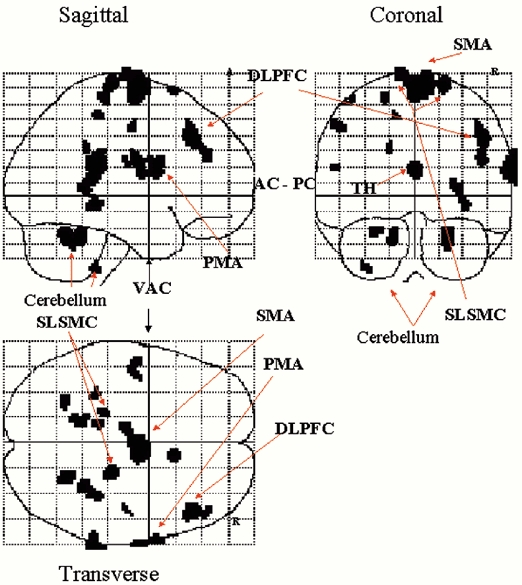
1. Positron emission tomography (PET) was used to identify the neuroanatomical correlates underlying 'central command' during imagination of exercise under hypnosis, in order to uncouple central command from peripheral feedback. 2. Three cognitive conditions were used: condition I, imagination of freewheeling downhill on a bicycle (no change in heart rate, HR, or ventilation, V(I)): condition II, imagination of exercise, cycling uphill (increased HR by 12 % and V(I) by 30 % of the actual exercise response): condition III, volitionally driven hyperventilation to match that achieved in condition II (no change in HR). 3. Subtraction methodology created contrast A (II minus I) highlighting cerebral areas involved in the imagination of exercise and contrast B (III minus I) highlighting areas activated in the direct volitional control of breathing (n = 4 for both; 8 scans per subject). End-tidal P(CO(2)) (P(ET,CO(2))) was held constant throughout PET scanning. 4. In contrast A, significant activations were seen in the right dorso-lateral prefrontal cortex, supplementary motor areas (SMA), the right premotor area (PMA), superolateral sensorimotor areas, thalamus, and bilaterally in the cerebellum. In contrast B, significant activations were present in the SMA and in lateral sensorimotor cortical areas. The SMA/PMA, dorso-lateral prefrontal cortex and the cerebellum are concerned with volitional/motor control, including that of the respiratory muscles. 5. The neuroanatomical areas activated suggest that a significant component of the respiratory response to 'exercise', in the absence of both movement feedback and an increase in CO(2) production, can be generated by what appears to be a behavioural response.


Figures

Figure 1
Figure 1. Imagination of exercise under hypnosis at rest
Imagination of heavy exercise for 2 min whilst under hypnosis (protocol 1) resulted in an increase in inspired air flow (top panel) and a decrease in PET,CO2 (bottom panel). Raw data from one subject. Filled bar indicates period of imagination of heavy exercise.
Figure 2
Figure 2. Imagination of heavy exercise under hypnosis
Group data for the imagination of heavy exercise under hypnosis (protocol 1, n = 17). Each data point represents the mean ± s.e.m. of a 15 s epoch, with the resting value (taken as the mean of the variable between 30 and 15 s prior to the onset of imagined exercise) subtracted from it. A, Δformula image; B, ΔPET,CO2; C, Δf; D, ΔVT; E, ΔHR (bpm, beats per minute).
Figure 3
Figure 3. Imagination of exercise under hypnosis at rest whilst maintaining isocapnia
Addition of CO2 to the inspired air to maintain isocapnia did not alter the magnitude of the ventilatory response to the imagination of exercise under hypnosis. Each data point represents the mean ± s.e.m. of a 15 s epoch, with the resting value (taken as the mean of the variable between 30 and 15 s prior to the onset of imagined exercise) subtracted from it (n = 7). A, Δformula image; B, ΔPET,CO2 (•, control; ○, isocapnic).
Figure 4
Figure 4. EMG activity during voluntary contraction and imagination of exercise
A comparison of voluntary leg movement (A) with the leg movement seen during imagination of exercise under hypnosis (B) in one subject. EMG recordings were made from left and right quadriceps and hamstrings. The vertical scale is the same in the two panels; the increase in EMG activity during imagination of exercise is minor.
Figure 5
Figure 5. Activations during imagination of exercise and voluntary hyperventilation with associated respiratory responses
Left, main activations during contrasts A and B rendered on the dorsal aspect of a representative brain in a transverse view from above (right edge, anterior) shown at P < 0.01 (uncorrected for multiple comparisons), for clarity. Only significant activations (Table 2) are labelled. Right, breath-by-breath ventilatory data in one subject for contrast A, imagining cycling uphill (ii), and for contrast B, breathing to an instructed frequency (iv) copying that in ii. Graphs i (contrast A) and iii (contrast B) record absence of effect when imagining freewheeling downhill. Each point represents a single breath and the 8 colours represent each repeat of the protocol. Experimental periods of 2 min 10 s are marked by the filled bars when scan performed. SMA, supplementary motor area; DLPFC, dorso-lateral prefrontal cortex. Activations within 2 cm of the brain surface are represented.
Figure 6
Figure 6. Areas of brain activated during imagination of exercise
Activations during imagination of exercise compared to imagination of freewheeling downhill (contrast A, P < 0.001, uncorrected for multiple comparisons) shown as through projections in standard stereotactic space (MNI). Only significant activations at P < 0.05 (Table 2) are labelled. R, right; A, anterior; AC-PC, commissural plane; VAC, vertical plane through anterior commissure; SMA, supplementary motor area; PMA, premotor area; DLPFC, dorso-lateral prefrontal cortex; TH, thalamus; SLSMC, superolateral sensorimotor cortex.
Figure 7
Figure 7. Transverse and sagittal activation during imagination of exercise
Two regions activated during imagination of exercise compared to imagination of freewheeling downhill, overlayed on a MRI. These areas are displayed at a level of P < 0.001 uncorrected for multiple comparisons. The Z statistic scale is shown in the lower right corner. The maximum activations in these areas, with Z > 4, are significant at P < 0.05 (Table 2). A, sagittal section (x = 4) showing activation of the SMA. B, transverse slice (z =−28) showing bilateral activation of the cerebellum.

References

    1. Adams L, Garlick J, Guz A, Murphy K, Semple SJ. Is the voluntary control of exercise in man necessary for the ventilatory response. Journal of Physiology. 1984;355:71–83. - PMC - PubMed
    1. Arvidsson T, Astrom H, Bevegard S, Jonsson B. Circulatory effects of suggested leg exercise and fear induced under hypnotic state. Progress in Respiration Research. 1970;5:365–374.
    1. Banner N, Guz A, Heaton R, Innes JA, Murphy K, Yacoub M. Ventilatory and circulatory responses at the onset of exercise in man following heart or heart-lung transplantation. Journal of Physiology. 1988;399:437–449. - PMC - PubMed
    1. Blessing WW. Inadequate frameworks for understanding bodily homeostasis. Trends in Neurosciences. 1997;20:235–239. - PubMed
    1. Coe W, St Jean R, Burger J. Hypnosis and the enhancement of visual imagery. International Journal of Clinical Experimental Hypnosis. 1980;28:225–243. - PubMed

Publication types