. 2001 Aug;13(4):199-212.
doi: 10.1002/hbm.1033.

A role for left temporal pole in the retrieval of words for unique entities

Affiliations

A role for left temporal pole in the retrieval of words for unique entities

T J Grabowski et al. Hum Brain Mapp. 2001 Aug.

Abstract

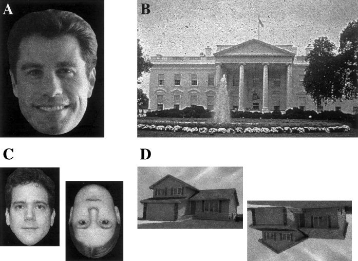
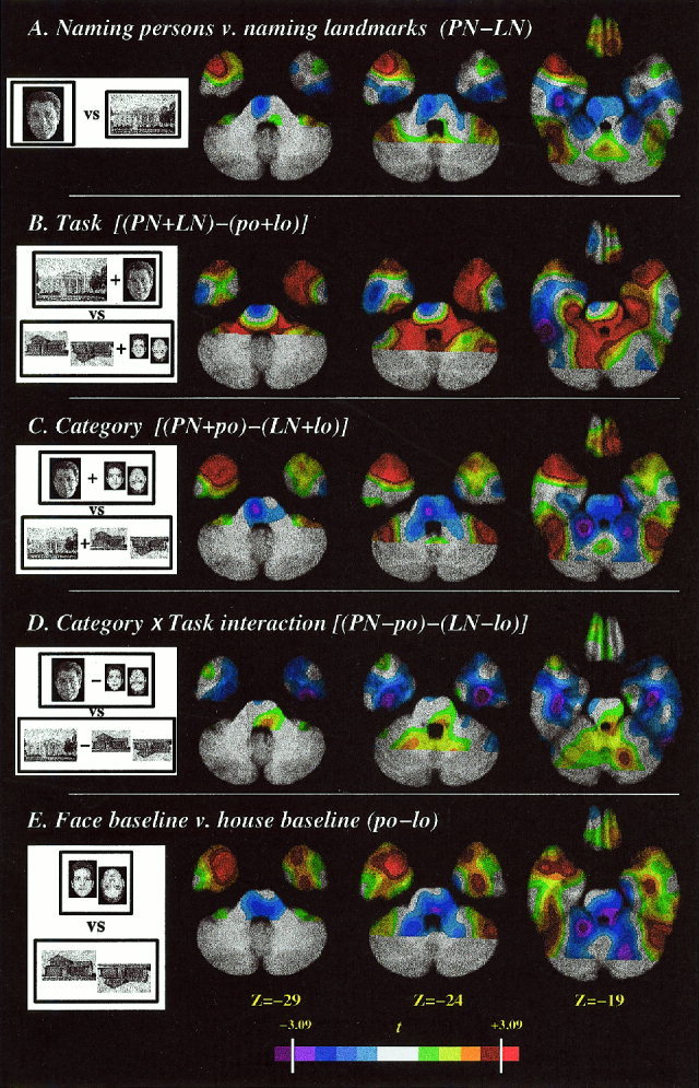
Both lesion and functional imaging studies have implicated sectors of high-order association cortices of the left temporal lobe in the retrieval of words for objects belonging to varied conceptual categories. In particular, the cortices located in the left temporal pole have been associated with naming unique persons from faces. Because this neuroanatomical-behavioral association might be related to either the specificity of the task (retrieving a name at unique level) or to the possible preferential processing of faces by anterior temporal cortices, we performed a PET imaging experiment to test the hypothesis that the effect is related to the specificity of the word retrieval task. Normal subjects were asked to name at unique level entities from two conceptual categories: famous landmarks and famous faces. In support of the hypothesis, naming entities in both categories was associated with increases in activity in the left temporal pole. No main effect of category (faces vs. landmarks/buildings) or interaction of task and category was found in the left temporal pole. Retrieving names for unique persons and for names for unique landmarks activate the same brain region. These findings are consistent with the notion that activity in the left temporal pole is linked to the level of specificity of word retrieval rather than the conceptual class to which the stimulus belongs.


Figures

Figure 1
Figure 1
Examples of experimental stimuli. A. Photographs of faces of famous persons. B. Photographs of famous buildings and natural landmarks. C. Photographs of faces of unknown persons. D. Photographs of unknown buildings.
Figure 2
Figure 2
Definition of the search volume. A. Upper left: The temporal pole was defined as the part of the temporal lobe anterior to a line (dotted yellow line) perpendicular to the long axis of the temporal lobe (red dotted line) at the level of the anterior ascending ramus of the Sylvian fissure. L and R ROIs were traced separately. Upper right: the three axial slices displayed in B. Lower left and right: 3D rendering of the average MR, with temporal poles, as defined for this study, painted red. B. Three Talairach axial sections, on which results are displayed in Figures 3 and 4. In the axial images, the right side of the image represents the left side of the brain.
Figure 3
Figure 3
T statistic maps for planned contrasts, superimposed on axial slices of the averaged MRI scan. The temporal pole search volume is delineated by the white dotted line (see Fig. 2 for details). The color scale at the bottom is calibrated with the significance level (3.09) determined by Gaussian random field theory. A. Naming persons contrasted with naming landmarks. B. Main effect of task (naming landmarks or persons contrasted with the baseline tasks). C. Main effect of category (naming or baseline using faces contrasted with naming or baseline using landmarks/buildings). D. Task by category interaction. (The difference between person naming and face baseline contrasted with the difference between landmark naming and building baseline.) E. Face baseline contrasted with building baseline. In the axial images, the right side of the image represents the left side of the brain.
Figure 4
Figure 4
T statistic map for the main effect of task, with the data smoothed with a 16‐mm kernel (top tier) or a 6‐mm kernel (bottom tier). The color scale is calibrated with the significance levels (3.09, 4.06, respectively) determined by Gaussian random field theory. The coordinates of the temporal polar activation (−41 +15 −19) discussed in the text are indicated on orthogonal views. In the axial images, the right side of the image represents the left side of the brain.
Figure 5
Figure 5
Left temporal polar activity (−41 +15 −19) across PET studies. This graph depicts PET activity, normalized to global activity of 1,000 counts, at the left temporal polar Talairach coordinate identified in Figure 4, across nine PET studies [including H. Damasio et al., 1996, 1999; Grabowski et al., 1998] including the one reported here, employing related tasks. Column 1: orientation decision baseline tasks with faces, ISI 1.0 sec, n = 6 studies; Column 2: orientation decision tasks using other concrete entities (unknown manmade objects, trees, or buildings), ISI 1.0 sec, n = 3. Column 3: orientation decision baseline tasks with faces, ISI 2.5 sec, n = 2; Column 4: orientation decision tasks using other concrete entities (unknown manmade objects, trees, or buildings), ISI 2.5 sec, n = 2; Column 5: retrieval of words for unique entities from this study and from Damasio et al. [1996], ISI 2.5 sec, n = 3. Column 6: Naming of manipulable manmade entities at basic object (n = 2) or subordinate level (n = 1), and verb generation (n = 1) for visually presented manipulable objects, ISI 2.5 sec. The normalized activity at this coordinate was greater in the three instances of unique level naming than it was in eight studies employing the face orientation control tasks (six with ISI 1 sec, two with ISI 2.5 sec); five studies employing orientation tasks with other classes of stimuli (buildings, trees, objects) at both 1 sec and 2.5 sec ISI; and four studies employing nonunique word production tasks using visual stimuli at an ISI of 2.5 sec (naming at basic object (2) and subordinate (1) level, and verb generation (1)). These data show that the activity at the left temporal polar coordinate in naming unique entities vs. baseline conditions is not simply a result of different ISIs for the retrieval and baseline tasks, as other retrieval tasks with the same ISI are not associated with relatively increased flow in the left temporal pole, and a reduced rate of performance of the baseline tasks is not associated with relatively increased flow in the left temporal pole.

References

    1. Aguirre GK, Zarahn E, D'Esposito M (1998a): An area within human ventral cortex sensitive to “building” stimuli: evidence and implications. Neuron 21: 373–383. - PubMed
    1. Aguirre GK, Zarahn E, D'Esposito M (1998b): Neural components of topographical representation. Proc Nat Acad Sci U S A 95: 839–846. - PMC - PubMed
    1. Albert MS, Butters N, Levin JA (1979): Temporal gradients in the retrograde amnesia of patients with alcoholic Korsakoff's disease. Arch Neurol 36: 211–216. - PubMed
    1. Allison TG, McCarthy G, Nobre A, Puce A, Belger A (1994): Human extrastriate visual cortex and the perception of faces, words, numbers, and colors. Cereb Cortex 5: 544–554. - PubMed
    1. Arnold SE, Hyman BT, Van Hoesen, GW (1994): Neuropathologic changes of the temporal pole in Alzheimer's disease and Pick's disease. Arch Neurol 51: 145–150. - PubMed

Publication types

LinkOut - more resources