. 2001 Jun;103(2):210-7.
doi: 10.1046/j.1365-2567.2001.01197.x.

Detection of anaphylatoxin receptors on CD83+ dendritic cells derived from human skin

Affiliations

Detection of anaphylatoxin receptors on CD83+ dendritic cells derived from human skin

K Kirchhoff et al. Immunology. 2001 Jun.

Abstract

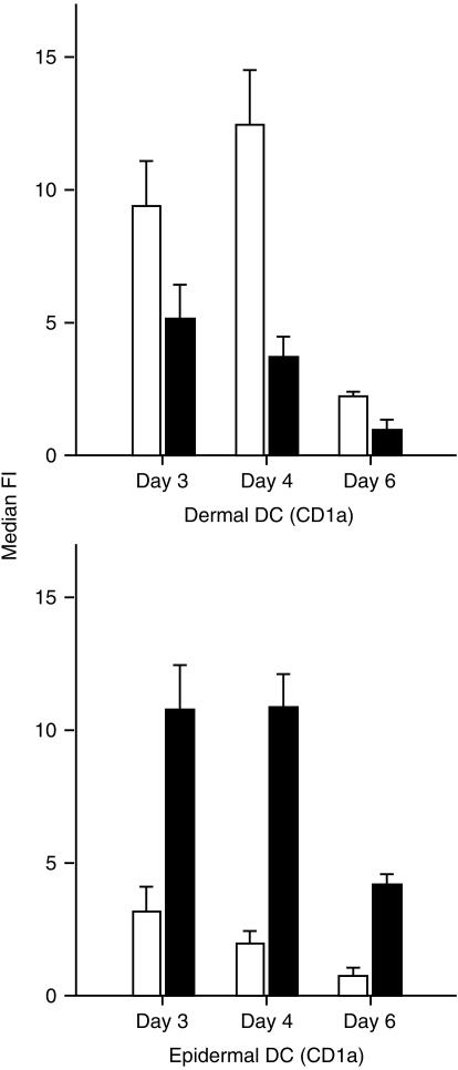
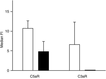
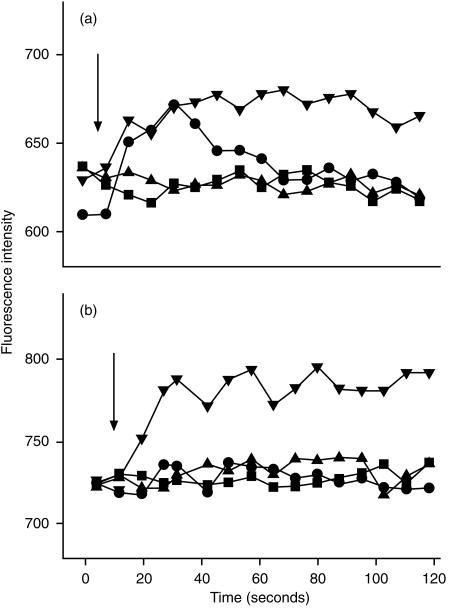
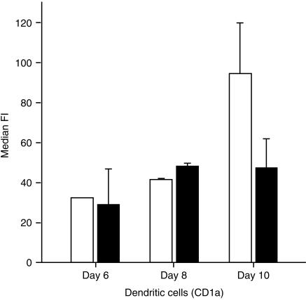
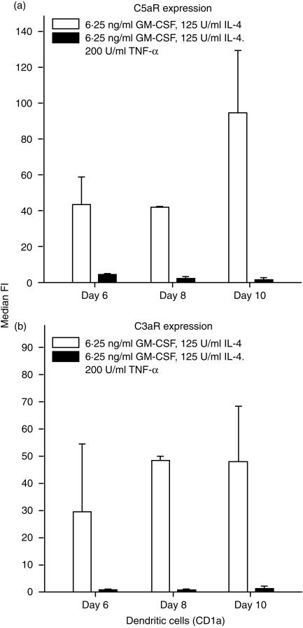
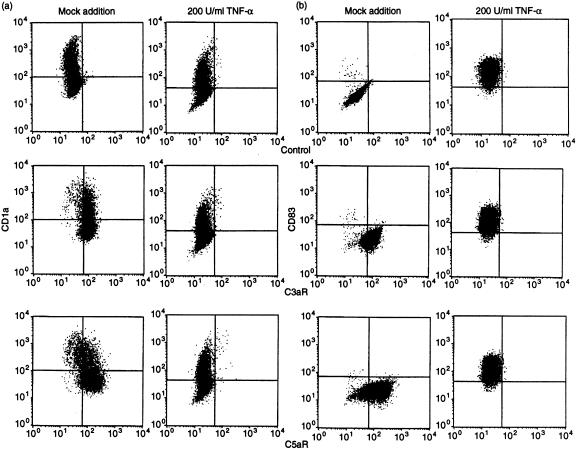
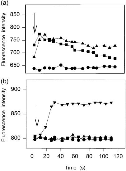
Dendritic cells (DC) are recruited to sites of inflammation for the initiation of immune responses. As the anaphylatoxins C5a and C3a are important mediators of inflammation, we investigated the expression of their receptors (C3aR and C5aR) on human DC. DC were isolated from human skin or generated from purified blood monocytes and were identified by their expression of CD1a or CD83. Freshly isolated or cultured dermal CD1a+ and CD83+ DC bound anti-C5aR and anti-C3aR monoclonal antibodies (mAbs), as detected by flow cytometry. C5a induced calcium fluxes in dermal CD1a+ and CD83+ DC, which could be inhibited by C17/5, an anti-C5a mAb. C3a did not induce calcium fluxes in these cells. Anaphylatoxin receptor expression was down-regulated on dermal DC by adding tumour necrosis factor-alpha (TNF-alpha) to the culture medium. On CD1a+ CD83- cells generated from isolated blood monocytes by culture with 6.25 ng/ml of granulocyte-macrophage colony-stimulating factor (GM-CSF) and 125 U/ml of interleukin-4 (IL-4), expression of both C5aR and C3aR was observed. In these cells, both C5a and C3a induced calcium fluxes. After addition of TNF-alpha to the culture medium, the majority of the CD1a+ cells expressed CD83+. These cells - expressing a phenotype of 'mature DC' - down-regulated the expression of the anaphylatoxin receptors and lost their reactivity to the respective ligands. Our results demonstrate the expression of the anaphylatoxin receptors C5aR and C3aR on human skin-derived DC and blood-derived cells expressing the DC-associated membrane molecule, CD1a. Furthermore, the expression of anaphylatoxin receptors on CD83+ dermal DC is indicative of an intermediate stage of maturation of these cells, which was not observed on in vitro-differentiated CD83+ cells.


Figures

Figure 1
Figure 1
Expression of receptors for C3a (▪) and C5a (□) on skin-derived dermal and epidermal dendritic cells (DC), which were cultured for up to 6 days. Dermal and epidermal cell suspensions were stained for simultaneous detection of anaphylatoxin receptors and CD1a, and analysed by flow cytometry. Results represent the average ±SD of four experiments. Only the median fluorescence intensity (median FI) of the CD1a+ cells is shown. Dermal DC express predominately C5aR, epidermal DC predominately C3aR.
Figure 2
Figure 2
Expression of receptors for C3a and C5a on skin-derived dendritic cells (DC), with (▪) and without (□) treatment with 200 U/ml of tumour necrosis factor-α (TNF-α). Dermal cell suspensions were cultured for 5 days and then stained for simultaneous detection of anaphylatoxin receptors and CD83 and analysed by flow cytometry. Results represent the average ±SD of four experiments. TNF-α decreases the expression of C5aR as well as of C3aR. Median FI, median fluorescence intensity.
Figure 3
Figure 3
Increase of cytosolic calcium in skin-derived dendritic cells (DC) upon stimulation with unlabelled C3a and C5a. Cells were cultured for 5 days (a). A proportion was treated with 200 U/ml of tumour necrosis factor-α (TNF-α) (b). After labelling with phycoerythrin-conjugated anti CD1a, DC were loaded with Flou-3 AM, as described in the Materials and methods. C3a (1 µg/ml) (▴) or C5a (1 µg/ml) (•) was then added to suspended cells and [Ca2+] was immediately assessed by flow cytometry. C5a was pretreated with a 20-fold molar excess of C17/5 (an anti-C5a monoclonal antibody [mAb]) in a control experiment (▪). Ca-ionophore was used as positive control (▾). C5a led to a transient calcium influx in C5aR+ dermal CD1a DC, which could be inhibited by preincubation with anti-C5a mAb (a). After stimulation with 200 U/ml of TNF-α, most of the cells became CD83+ and no longer showed calcium influx (b). Arrows refer to the time point when C3a resp. C5a were added to the cell suspension.
Figure 4
Figure 4
Expression of receptors for C3a (▪) and C5a (□) on blood-derived dendritic cells, which were cultured in the presence of interleukin-4 (IL-4) (125 U/ml) and granulocyte–macrophage colony-stimulating factor (GM-CSF) (6·25 ng/ml) for up to 10 days. Cells were stained for simultaneous detection of anaphylatoxin receptors and CD1a, and analysed by flow cytometry. Results represent the average ±SD of three experiments. Only the median fluorescence intensity (median FI) of the CD1a+ cells is shown. Blood-derived dendritic cells express both C5aR and C3aR.
Figure 5
Figure 5
Expression of receptors for C3a and C5a on blood-derived cells, with (▪) and without (□) treatment with 200 U/ml of tumour necrosis factor-α (TNF-α). Cell suspensions were cultured for 6, 8 or 10 days and then stained for the simultaneous expression of anaphylatoxin receptors and CD1a, and analysed by flow cytometry. Results represent the average ±SD of three experiments. TNF-α inhibits the expression of both C5aR and C3aR. GM-CSF, granulocyte–macrophage colony-stimulating factor.
Figure 6
Figure 6
Changes in the expression of anaphylatoxin receptors on blood-derived cells following treatment with 200 U/ml of tumour necrosis factor-a (TNF-α). Cells were cultured for 5 days and then processed for double staining using monoclonal antibodies (mAbs) specific for C3aR and C5aR. The presence of C3aR and C5aR was revealed by incubation with fluorescein isothiocyanate (FITC)-conjugated anti-mouse immunoglobulins followed by phycoerythrin-conjugated anti-CD1a (a) or anti-CD83 (b). Both CD1a+ and CD1a cells showed expression of C3aR and C5aR, which was completely down-modulated by treatment with TNF-α (a). The treatment of these cells with TNF-α led to the expression of CD83 and to the disappearance of the anaphylatoxin receptors (b).
Figure 7
Figure 7
Increase of cytosolic calcium in blood-derived cells upon stimulation with unlabelled C3a and C5a. Cells were cultured for 5 days (a). A proportion was then stimulated with 200 U/ml of tumour necrosis factor-α (TNF-α) (b). After cell labelling with phycoerythrin-conjugated anti-CD1a, cells were loaded with Flou-3 AM, as described in the Materials and methods. C3a (1 µg/ml) (▴) or C5a (1 µg/ml) (•) was then added to suspended cells and [Ca2+] was immediately assessed by flow cytometry. Ca-ionophore was used as a positive control (▾) and buffer as a negative control (▪). Cytosolic calcium increased in blood-derived cells after stimulation with unlabelled C3a and C5a (a). After stimulation with 200 U/ml of TNF-α, all cells became CD83+ and a calcium influx was no longer observed (b).

References

    1. Bos JD, Kapsenberg ML. The skin immune system: progress in cutaneous biology. Immunol Today. 1993;14:75–8. - PubMed
    1. Cella M, Scheidegger D, Palmer-Lehmann K, Lane P, Lanzavecchia A, Alber G. Ligation of CD40 on dendritic cells triggers production of high levels of interleukin-12 and enhances T cell stimulatory capacity: T–T help via APC activation. J Exp Med. 1996;184:747–52. - PMC - PubMed
    1. Koch F, Stanzl U, Jennewein P, Janke K, Heufler C, Kampgen E, Romani N, Schuler G. High level IL-12 production by murine dendritic cells: upregulation via MHC class II and CD40 molecules and downregulation by IL-4 and IL-10. J Exp Med. 1996;184:741–6. - PMC - PubMed
    1. Steinman RM. The dendritic cell system and its role in immunogenicity. Annu Rev Immunol. 1991;9:271–96. - PubMed
    1. Caux C, Liu YJ, Banchereau J. Recent advances in the study of dendritic cells and follicular dendritic cells. Immunol Today. 1995;16:2–4. - PubMed

Publication types

MeSH terms