. 2001 Jul;49(1):73-81.
doi: 10.1136/gut.49.1.73.

LI-cadherin: a marker of gastric metaplasia and neoplasia

Affiliations

LI-cadherin: a marker of gastric metaplasia and neoplasia

C Grötzinger et al. Gut. 2001 Jul.

Abstract

Background: Intestinal metaplasia is considered a risk factor for the development of gastric adenocarcinomas of the intestinal type and is found in approximately 20% of gastric biopsies. Conventional histology only detects advanced stages of intestinal metaplasia.

Aims: To study expression of the enterocyte specific adhesion molecule liver-intestinal (LI)-cadherin in intestinal metaplasia as well as in gastric cancer, and to evaluate its use as a diagnostic marker molecule.

Patients: Gastric biopsies (n=77) from 30 consecutive patients (n=30; aged 28-90 years) as well as surgically resected tissue samples (n=24) of all types of gastric carcinomas were analysed.

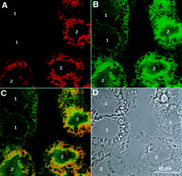
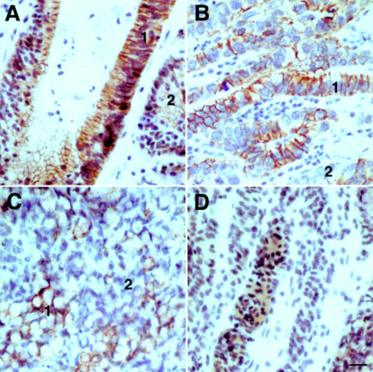
Methods: Single and double label immunofluorescence detection on cryosections of gastric biopsies; alkaline phosphatase antialkaline phosphatase method on paraffin embedded carcinoma tissue sections.

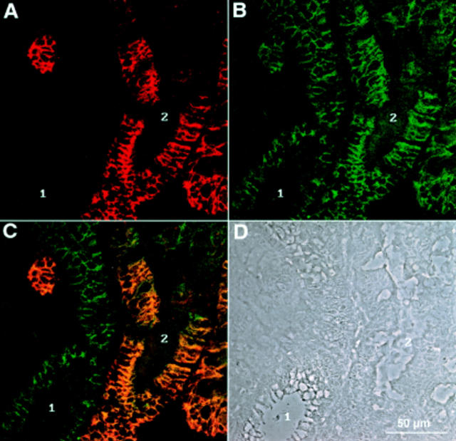
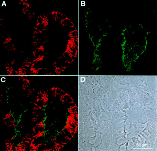
Results: Of 77 biopsies (from 30 patients), 12 (from 10 patients) stained positive for LI-cadherin. LI-cadherin staining correlated with the presence of intestinal metaplasia. Conventional histological diagnosis however failed to detect subtle gastric intestinal metaplasia (three of 10 patients). In contrast, only LI-cadherin and villin were positive in these cases whereas sucrase-isomaltase also failed to detect intestinal metaplasia in four of 10 patients. Well differentiated gastric carcinomas showed intense staining for LI-cadherin while undifferentiated carcinomas showed only weak diffuse cytoplasmic staining.

Conclusions: To detect early metaplastic changes in the gastric mucosa, LI-cadherin has a sensitivity superior to sucrase-isomaltase and conventional histology and comparable with that of villin. Its specificity exceeds that of villin. Thus LI-cadherin represents a new, reliable, and powerful marker molecule for early detection of gastric intestinal metaplasia and well differentiated adenocarcinomas.


Figures

Figure 1
Figure 1
Western blot analysis of LI-cadherin expression in the upper gastrointestinal tract. Protein extracts of biopsies (oesophagus, stomach, duodenum, ileum, and jejunum) and of the human colon carcinoma cell line Caco-2 were separated by sodium dodecyl sulphate-polyacrylamide gel electrophoresis and immunoblotted with polyclonal antiserum against human LI-cadherin.
Figure 2
Figure 2
Immunofluorescence microscopy of a gastric biopsy stained with polyclonal antibody against LI-cadherin: (A) fluorescence signal of LI-cadherin, (B) phase contrast. Note the distinct LI-cadherin staining of goblet cells in one gland whereas surrounding glands are negative.
Figure 3
Figure 3
Double immunofluorescence recorded by confocal laser microscopy of a gastric biopsy stained with polyclonal antiserum against LI-cadherin (A) and monoclonal antibody against E-cadherin (B). (C) Combined images of both proteins. The phase contrast image of the same area (D) reveals regions of normal mucosa (1) next to those showing characteristics of intestinal metaplasia (2). Note the high degree of colocalisation in cells expressing both LI-cadherin and E-cadherin.
Figure 4
Figure 4
Double immunofluorescence recorded by confocal laser microscopy of a gastric biopsy with polyclonal antiserum against LI-cadherin (A) and monoclonal antibody against sucrase-isomaltase (B). (C) Combined images of both proteins. (D) Phase contrast image of the same area. Whereas LI-cadherin expression is restricted to the basolateral plasma membrane regions, sucrase-isomaltase is exclusively found in the apical regions and does not colocalise with LI-cadherin.
Figure 5
Figure 5
Immunofluorescence recorded by confocal laser microscopy of a gastric biopsy with polyclonal antiserum against LI-cadherin (A) and monoclonal antibody against villin (B). (C) Combined images of both proteins. (D) Phase contrast image of the same area. Whereas LI-cadherin background is very low in non-metaplastic regions (1, A), villin staining results typically in higher background staining (1, B). Connective tissue in between the glands is totally negative for both proteins (centre).
Figure 6
Figure 6
Immunoreactivity of LI-cadherin in gastric carcinomas. Immunohistochemical staining of LI-cadherin in mucinous (A), tubular (B), and signet ring (C) adenocarcinoma, and in undifferentiated carcinoma of the stomach (D). Note the intense staining of the cell-cell contact areas in well differentiated regions (1) of adenocarcinomas contrasted by weaker staining in less differentiated regions (2). The undifferentiated carcinoma (D) exhibits only faint cytoplasmic staining for LI-cadherin.

Comment in

References

    1. Gut. 1985 Dec;26(12):1319-26 - PubMed
    1. J Cell Biol. 1985 Sep;101(3):838-51 - PubMed
    1. Gastroenterology. 1987 Jan;92(1):98-105 - PubMed
    1. Virchows Arch B Cell Pathol Incl Mol Pathol. 1987;54(3):155-69 - PubMed
    1. Virchows Arch A Pathol Anat Histopathol. 1988;413(4):303-12 - PubMed

MeSH terms