

Tumour necrosis factor alpha impairs function of liver derived T lymphocytes and natural killer cells in patients with primary sclerosing cholangitis
- PMID: 11413121
- PMCID: PMC1728361
- DOI: 10.1136/gut.49.1.131
Tumour necrosis factor alpha impairs function of liver derived T lymphocytes and natural killer cells in patients with primary sclerosing cholangitis
Abstract
Background: Primary sclerosing cholangitis (PSC) is considered to be a chronic autoimmune disease where infiltrating T lymphocytes have been implicated in the destruction of bile ducts. Altered function of these T cells may reflect abnormalities in the immune response leading to tissue damage.
Aim: We investigated the proliferative and functional capacity of freshly isolated liver derived T lymphocytes (LDLs) and natural killer (NK) cells from PSC patients.
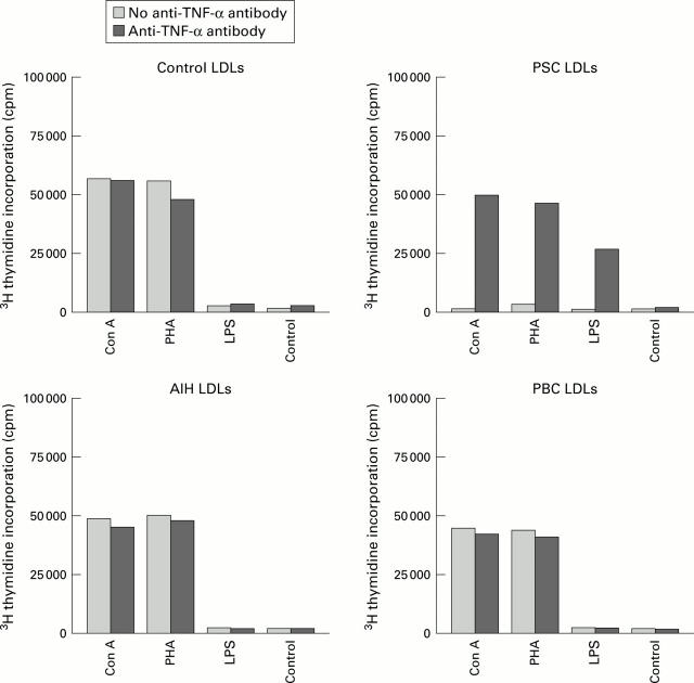
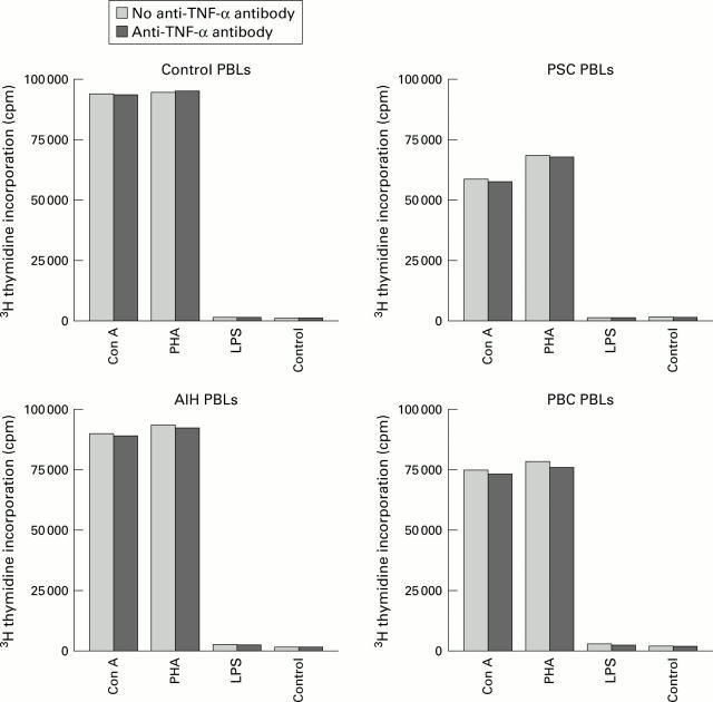
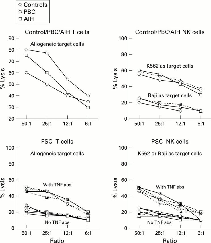
Methods: The proliferative responses to common mitogens such as phytohaemagglutinin (PHA), concanavalin A (Con A), and lipopolysaccharide (LPS) were studied, and the cytotoxic function of T lymphocytes was measured using allogeneic target cells. NK (CD56(+)/16(+)) cytotoxic function was measured using the two cell lines K562 (NK sensitive) and Raji lymphoma cells (NK resistant).
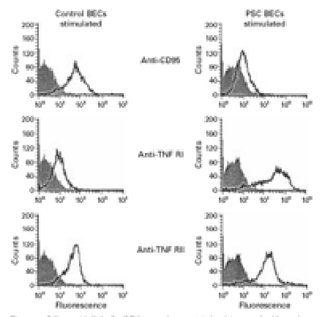
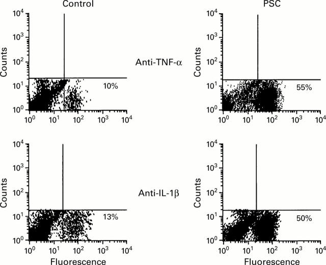
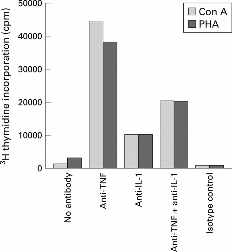
Results: Compared with patients with primary biliary cirrhosis (PBC), autoimmune hepatitis (AIH), and normal controls (without liver disease), in PSC: (1) LDLs contained a low percentage of T cells; (2) there was significantly decreased expression of interleukin (IL)-2 receptor (p<0.001) on activated T cells (HLA-DR(+)); (3) LDLs but not peripheral blood lymphocytes had significantly impaired proliferative responses to mitogens such as PHA, Con A, and LPS (p< 0.001); (4) no cytotoxic activity of PSC liver T and NK cells was recorded; (5) significantly higher levels of tumour necrosis factor alpha (TNF-alpha) and IL-1beta but lower levels of IL-2, IL-10, and interferon gamma were found in the supernatants of mitogen stimulated LDL cultures (p<0.001); (6) higher percentages of freshly isolated PSC LDLs contained intracytoplasmic TNF-alpha and IL-1beta; and (7) pretreatment of PSC LDLs in vitro with neutralising TNF antibodies significantly enhanced proliferative responses and allowed IL-2 receptor expression following stimulation. In addition, the impaired cytolytic activity of both NK and T cells was partially restored. Impaired proliferative or functional capacity of liver derived T cells was not observed in either PBC or AIH patients.
Conclusions: We suggest that reduced T cell reactivity in liver infiltrating cells obtained from patients with PSC is due to high local production of TNF-alpha. Our findings indicate that the use of anti-TNF antibodies as an alternative treatment for PSC patients should be evaluated.
Figures
References
Publication types
MeSH terms
Substances
LinkOut - more resources
Full Text Sources
Research Materials