. 2001 Jun;107(12):1529-36.
doi: 10.1172/JCI12568.

Transient expression of IL-1beta induces acute lung injury and chronic repair leading to pulmonary fibrosis

Affiliations

Transient expression of IL-1beta induces acute lung injury and chronic repair leading to pulmonary fibrosis

M Kolb et al. J Clin Invest. 2001 Jun.

Abstract

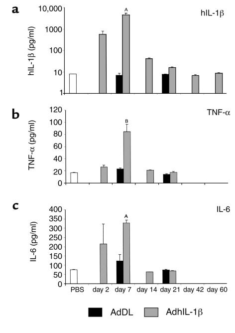
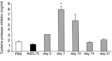
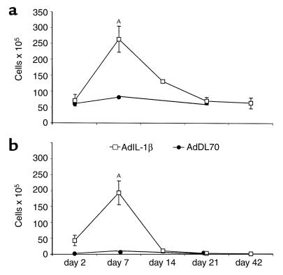
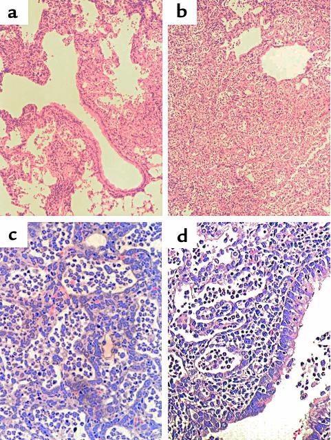
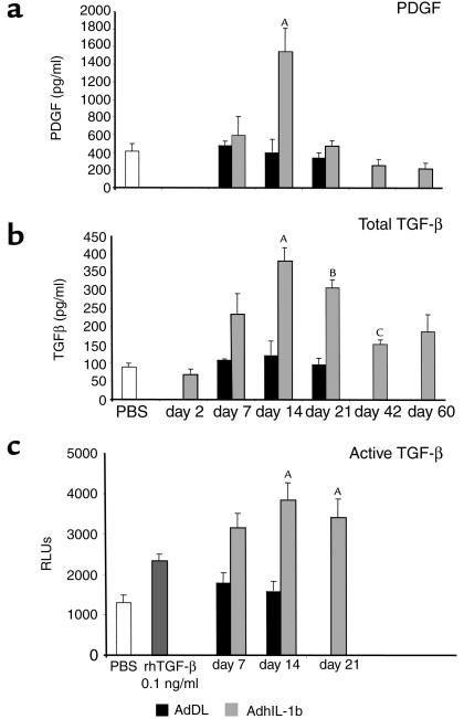
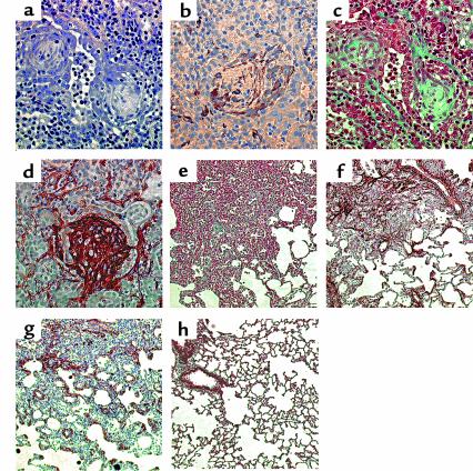
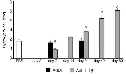
IL-1beta is one of a family of proinflammatory cytokines thought to be involved in many acute and chronic diseases. Although it is considered to participate in wound repair, no major role has been attributed to IL-1beta in tissue fibrosis. We used adenoviral gene transfer to transiently overexpress IL-1beta in rat lungs after intratracheal administration. The high expression of IL-1beta in the first week after injection was accompanied by local increase of the proinflammatory cytokines IL-6 and TNF-alpha and a vigorous acute inflammatory tissue response with evidence of tissue injury. The profibrotic cytokines PDGF and TGF-beta1 were increased in lung fluid samples 1 week after peak expression of IL-1beta. Although PDGF returned to baseline in the third week, TGF-beta1 showed increased concentrations in bronchoalveolar lavage fluid for up to 60 days. This was associated with severe progressive tissue fibrosis in the lung, as shown by the presence of myofibroblasts, fibroblast foci, and significant extracellular accumulations of collagen and fibronectin. These data directly demonstrate how acute tissue injury in the lung, initiated by a highly proinflammatory cytokine, IL-1beta, converts to progressive fibrotic changes. IL-1beta should be considered a valid target for therapeutic intervention in diseases associated with fibrosis and tissue remodeling.


Figures

Figure 1
Figure 1
Proinflammatory cytokines in BAL fluid. (a) Transgene human IL-1β was elevated 2 days after injection of AdhIL-1β, was maximally expressed after 7 days, and returned to baseline by day 14. Endogenous TNF-α (b) and IL-6 (c) were upregulated parallel to hIL-1β and returned to baseline concentration by day 14. In control animals treated with AdDL70, hIL-1β, TNF-α, and IL-6 were not different from PBS-treated rats. AP < 0.008 versus AdDL70 control; BP < 0.02 versus AdDL70 control.
Figure 2
Figure 2
Serum concentration of α1-cysteine protease inhibitor was maximally increased 7 days after injection of AdhIL-1β and returned to baseline by day 14. Injection of control virus AdDL70 did not cause elevated serum levels of α1-cysteine protease inhibitor. AP < 0.002 versus AdDL70 control.
Figure 3
Figure 3
(a) Total cells retrieved by BAL were increased 7 and 14 days after injection of AdhIL-1β. The vast majority of cells at day 7 were neutrophils (b), whereas alveolar macrophages accounted for the persistent increase of total cells at day 14. Cell counts and differentials were normal by day 21. Seven days after injection, animals treated with control virus AdDL70 had a slight, insignificant increase in total cell number. Neutrophils were not elevated. AP < 0.01 versus AdDL70 control.
Figure 4
Figure 4
Lung histology in the first week after injection of AdhIL-1β showed acute inflammatory reactions in the lungs, beginning in peribronchial areas after 2 days (a). By day 7, inflammatory infiltrates affected large parts of the lung (b). In some areas, the cellular infiltrates completely filled alveoli while alveolar structures remained intact. In other regions, they caused marked alveolar wall destruction (c). The inflammatory infiltrates consisted mainly of neutrophils and macrophages, reaching the airway lumen after passing bronchial mucosa (d). Hematoxylin and eosin staining; ×24 (a and b); ×160 (c and d).
Figure 5
Figure 5
Profibrotic cytokines in BAL fluid. (a) PDGF was increased 2 weeks after injection of AdhIL-1β, 1 week after peak expression of proinflammatory cytokines, and returned to normal values by day 21. (b) TGF-β also showed maximal expression after 2 weeks. By day 21, in contrast to PDGF, concentration of TGF-β in BAL fluid was slightly reduced compared with day 14. Increased TGF-β concentration was measured for the entire course of the experiment up to day 60. (c) The presence of bioactive TGF-β in BAL fluids of AdhIL-1β–treated animals was demonstrated using an established bioassay (MLECs with luciferase reporter gene, controlled by TGF-β–inducible promoter PAI-1; for details see the text). AP < 0.04 versus AdDL70 control. BP < 0.02 versus AdDL70 control. CP < 0.04 versus PBS.
Figure 6
Figure 6
Fibrogenic responses in the lung after intratracheal injection of AdhIL-1β. (a) Numerous fibroblast foci were observed in the lung tissue 14 days after injection. Special staining methods demonstrated that cells in these foci were myofibroblasts (b, α-SMA immunohistochemistry), and synthesized collagen (c, Masson trichrome) and fibronectin (d, immunohistochemistry). Major collagen and fibronectin accumulation was noticed 3 weeks after injection of AdhIL-1β (e and f, Masson trichrome and fibronectin immunohistochemistry), and myofibroblasts were present in interstitial areas and along alveolar walls (g, α-SMA immunohistochemistry). The histological appearance of animals treated with control virus AdDL70 was normal (h, Masson trichrome). ×160 (ad); ×50 (eh).
Figure 7
Figure 7
Animals treated with AdhIL-1β showed a progressive increase of lung hydroxyproline concentration past day 21 after injection. AP < 0.01 versus AdDL70 control. BP < 0.01, CP < 0.001 versus PBS control.

Comment in

References

    1. Dinarello CA. Interleukin-1. Cytokine Growth Factor Rev. 1997;8:253–265. - PubMed
    1. Tocci, M.J., and Schmidt, J.A. 1997. Interleukin-1: structure and function. In Cytokines in health and disease. D.G. Remick and J.S. Friedland, editors. Marcel Dekker. New York, New York, USA. 1–27.
    1. Watkins LR, Hansen MK, Nguyen KT, Lee JE, Maier SF. Dynamic regulation of the proinflammatory cytokine, interleukin-1beta: molecular biology for non-molecular biologists. Life Sci. 1999;65:449–481. - PubMed
    1. Phan SH, Kunkel SL. Lung cytokine production in bleomycin-induced pulmonary fibrosis. Exp Lung Res. 1992;18:29–43. - PubMed
    1. Thrall, R.S., and Scalise, P.J. 1995. Bleomycin. In Pulmonary fibrosis. S.H. Phan, editor. Marcel Dekker. New York, New York, USA. 231–292.

Publication types

MeSH terms