. 2001 Jun 18;193(12):1361-71.
doi: 10.1084/jem.193.12.1361.

Role of promyelocytic leukemia (PML) sumolation in nuclear body formation, 11S proteasome recruitment, and As2O3-induced PML or PML/retinoic acid receptor alpha degradation

Affiliations

Role of promyelocytic leukemia (PML) sumolation in nuclear body formation, 11S proteasome recruitment, and As2O3-induced PML or PML/retinoic acid receptor alpha degradation

V Lallemand-Breitenbach et al. J Exp Med. .

Abstract

Promyelocytic leukemia (PML) is the organizer of nuclear matrix domains, PML nuclear bodies (NBs), with a proposed role in apoptosis control. In acute promyelocytic leukemia, PML/retinoic acid receptor (RAR) alpha expression disrupts NBs, but therapies such as retinoic acid or arsenic trioxide (As2O3) restore them. PML is conjugated by the ubiquitin-related peptide SUMO-1, a process enhanced by As2O3 and proposed to target PML to the nuclear matrix. We demonstrate that As2O3 triggers the proteasome-dependent degradation of PML and PML/RARalpha and that this process requires a specific sumolation site in PML, K160. PML sumolation is dispensable for its As2O3-induced matrix targeting and formation of primary nuclear aggregates, but is required for the formation of secondary shell-like NBs. Interestingly, only these mature NBs harbor 11S proteasome components, which are further recruited upon As2O3 exposure. Proteasome recruitment by sumolated PML only likely accounts for the failure of PML-K160R to be degraded. Therefore, studying the basis of As2O3-induced PML/RARalpha degradation we show that PML sumolation directly or indirectly promotes its catabolism, suggesting that mature NBs could be sites of intranuclear proteolysis and opening new insights into NB alterations found in viral infections or transformation.


Figures

Figure 1
Figure 1
Arsenic induces SUMO modification and proteasome-dependant degradation of PML. (a) Western blot analysis of His-purified proteins from His-PML-CHO cells treated or not with 1 μM As2O3 overnight and revealed with anti-PML antibodies (left), with anti–SUMO-1 antibodies (middle), or anti–SUMO-2 antibodies (right). (b) Western blot analysis performed on whole CHO–PML extract revealed with anti-PML and anti-actin antibodies. Cells were treated overnight with 1 μM As2O3, 10 μM lactacystin (L), both (L/As), or none (φ). SUMO-modified PML species are indicated. (c) HeLa cells were treated for 48 h with IFN-α or -γ, with or without an overnight As2O3 treatment. PML isoforms induced by IFN are degraded after As2O3 treatment (top), whereas Sp100 isoforms are not degraded (bottom). (d) PML–CHO cells were treated or not with 1 μM As2O3 and 10 nM LMB for 12 h.
Figure 2
Figure 2
The COOH terminal part of PML proteins is required for their As2O3-induced degradation. (a) Schematic representation of As2O3-induced degradation of different PML mutants and another isoform (PML-4). (b) Western blot analysis of some of the mutants depicted above with or without an overnight 1 μM As2O3 treatment. Unsumolated PML proteins are indicated by arrows. PML mutants were stably expressed in CHO cells from pSG5 expression vectors.
Figure 2
Figure 2
The COOH terminal part of PML proteins is required for their As2O3-induced degradation. (a) Schematic representation of As2O3-induced degradation of different PML mutants and another isoform (PML-4). (b) Western blot analysis of some of the mutants depicted above with or without an overnight 1 μM As2O3 treatment. Unsumolated PML proteins are indicated by arrows. PML mutants were stably expressed in CHO cells from pSG5 expression vectors.
Figure 3
Figure 3
SUMO modification of K160 is required for As2O3-induced PML and PML/RARα degradation. (a) When stably expressed in CHO cells, PML-3K is not degraded upon an overnight As2O3 (As) exposure but upregulated by lactacystin (L) treatment. (b) SUMO modification of K160R and K490R PML mutants stably overexpressed in CHO treated with 1 μM As2O3 for 1 h were analyzed by Western blot analysis. (c) A longer As2O3 exposure (12 h) induces degradation of wild-type PML and K490R mutant, but not K160R. (d) Mutation of K160 in PML/RARα abolishes As2O3-induced degradation (12-h treatment) in retrovirally transduced NIH3T3 cells. This Western blot was revealed with an anti-RARα antibody.
Figure 3
Figure 3
SUMO modification of K160 is required for As2O3-induced PML and PML/RARα degradation. (a) When stably expressed in CHO cells, PML-3K is not degraded upon an overnight As2O3 (As) exposure but upregulated by lactacystin (L) treatment. (b) SUMO modification of K160R and K490R PML mutants stably overexpressed in CHO treated with 1 μM As2O3 for 1 h were analyzed by Western blot analysis. (c) A longer As2O3 exposure (12 h) induces degradation of wild-type PML and K490R mutant, but not K160R. (d) Mutation of K160 in PML/RARα abolishes As2O3-induced degradation (12-h treatment) in retrovirally transduced NIH3T3 cells. This Western blot was revealed with an anti-RARα antibody.
Figure 4
Figure 4
SUMO modification of PML is dispensable for PML matrix transfer induced by As2O3. (a) CHO–PML were treated with As2O3 for 1 h (As1h), for 12 h (As), and/or with lactacystin for 12 h (L and L/As). Proteins were fractionated into RIPA (R) or pellet (P) fractions before Western blot analysis (lanes are indicated). (b–d) PML−/− MEF stably transfected wild-type PML or PML-3K mutant were analyzed after a 1-h As2O3 treatment. (b) Note the As2O3-induced matrix transfer of PML-3K. (c) As2O3-induced matrix targeting of PML-3K is blocked by pretreatment with okadaic acid. Okadaic acid induces an additional, likely a phosphorylated, PML species. (d) Immunofluorescence analysis of in situ nuclear matrix preparation from PML−/− cells stably expressing wild-type PML or PML-3K mutant. Note the As2O3-induced aggregation of NBs for PML, but not for the PML-3K mutant, which remains in distinctly smaller aggregates.
Figure 4
Figure 4
SUMO modification of PML is dispensable for PML matrix transfer induced by As2O3. (a) CHO–PML were treated with As2O3 for 1 h (As1h), for 12 h (As), and/or with lactacystin for 12 h (L and L/As). Proteins were fractionated into RIPA (R) or pellet (P) fractions before Western blot analysis (lanes are indicated). (b–d) PML−/− MEF stably transfected wild-type PML or PML-3K mutant were analyzed after a 1-h As2O3 treatment. (b) Note the As2O3-induced matrix transfer of PML-3K. (c) As2O3-induced matrix targeting of PML-3K is blocked by pretreatment with okadaic acid. Okadaic acid induces an additional, likely a phosphorylated, PML species. (d) Immunofluorescence analysis of in situ nuclear matrix preparation from PML−/− cells stably expressing wild-type PML or PML-3K mutant. Note the As2O3-induced aggregation of NBs for PML, but not for the PML-3K mutant, which remains in distinctly smaller aggregates.
Figure 5
Figure 5
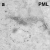
SUMO modification of PML is required for mature NBs formation. Electronic microscopy examination of PML−/− MEF stably expressing PML (a) or PML-3K mutant (b). Note that small dense aggregates are formed by PML-3K, whereas standard PML empty structures where PML forms a distinct outer rim are observed upon expression of wild-type PML (reference 14).
Figure 5
Figure 5
SUMO modification of PML is required for mature NBs formation. Electronic microscopy examination of PML−/− MEF stably expressing PML (a) or PML-3K mutant (b). Note that small dense aggregates are formed by PML-3K, whereas standard PML empty structures where PML forms a distinct outer rim are observed upon expression of wild-type PML (reference 14).
Figure 7
Figure 7
PML-3K still recruits CBP, but not Daxx or Sp100, while PML-K160R fails to recruit 11Sα proteasome upon As2O3 exposure. Immuno-fluorescences were performed on PML−/− MEFs transiently transfected with wild-type PML or PML-3K mutant, stained with anti-PML, anti-Sp100, anti-Daxx, and monoclonal anti-CBP antibodies (top). PML−/− MEF cells infected with a PML or PML-K160R–expressing retrovirus were treated with As2O3 for 1 h and stained with PML or anti-11Sα antibodies as indicated (bottom). Note that 11Sα does not colocalize with PML-K160R.
Figure 6
Figure 6
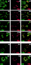
11S proteasome is recruited by PML onto NBs. Confocal analysis of PML and various proteasome components were realized on PML overexpressing CHO treated for 1 h by As2O3 (As), lactacystin (L), none (φ), or both (L/As). Localization of the endogenous 20S core and 11Sα, or β regulatory subunits of the proteasome are compared with that of PML as indicated.
Figure 8
Figure 8
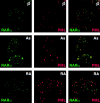
As2O3 targets PML/RARα onto NBs during As2O3-induced but not RA-induced degradation. NB4 cells were exposed to 1 μM As2O3 or RA for 6 and 24 h, respectively, before immunofluorescence, as indicated.
Figure 9
Figure 9
Schematic representation of PML traffic onto NBs. Under our working model, PML is initially dispersed in the nucleoplasm possibly in the chromatin. A specific dephosphorylation event triggered by As2O3 targets PML to the nuclear matrix on primary PML bodies. Sumolation then induces the maturation to secondary PML bodies that contain the 11S proteasome subunits α and β, Daxx, and Sp100, where PML would be degraded.

References

    1. de Thé H., Lavau C., Marchio A., Chomienne C., Degos L., Dejean A. The PML-RARα fusion mRNA generated by the t(15;17) translocation in acute promyelocytic leukemia encodes a functionally altered RAR. Cell. 1991;66:675–684. - PubMed
    1. Freemont P.S. RING for destruction? Curr. Biol. 2000;10:R84–R87. - PubMed
    1. Wang Z.-G., Ruggero D., Ronchetti S., Zhong S., Gaboli M., Rivi R., Pandolfi P.P. PML is essential for multiple apoptotic pathways. Nat. Genet. 1998;20:266–272. - PubMed
    1. Wang Z.G., Delva L., Gaboli M., Rivi R., Giorgio M., Cordon-Cardo C., Grosveld F., Pandolfi P.P. Role of PML in cell growth and the retinoic acid pathway. Science. 1998;279:1547–1551. - PubMed
    1. Quignon F., de Bels F., Koken M., Feunteun J., Ameisen J.-C., de Thé H. PML induces a caspase-independent cell death process. Nat. Genet. 1998;20:259–265. - PubMed

Publication types

MeSH terms