. 2001 Jun 18;193(12):1425-30.
doi: 10.1084/jem.193.12.1425.

A transcription function for the T cell-specific adapter (TSAd) protein in T cells: critical role of the TSAd Src homology 2 domain

Affiliations

A transcription function for the T cell-specific adapter (TSAd) protein in T cells: critical role of the TSAd Src homology 2 domain

F Marti et al. J Exp Med. .

Abstract

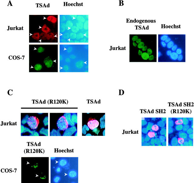
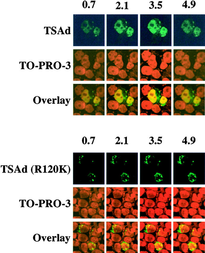
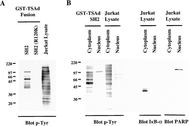
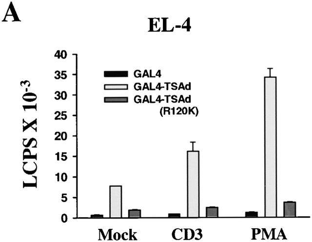
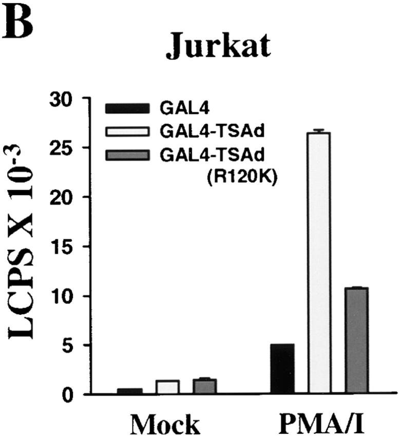
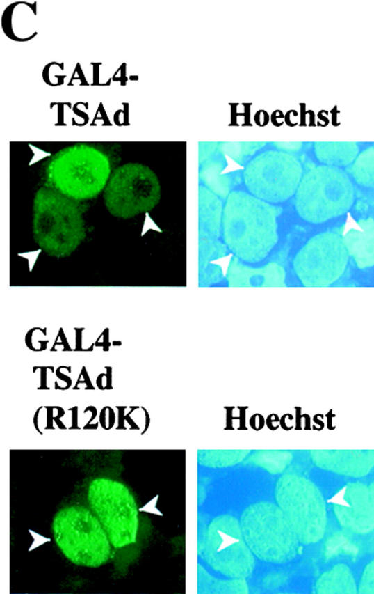
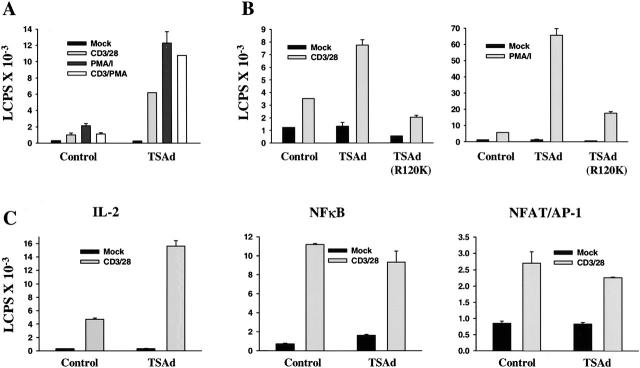
T cell-specific adapter (TSAd) protein is an Src homology 2 (SH2) domain-containing adapter molecule implicated in T cell receptor for antigen (TCR)-mediated interleukin 2 (IL-2) secretion in T cells. Here, we demonstrate that a substantial fraction of TSAd is found in the T cell nucleus. Nuclear import of TSAd is an active process that depends on TSAd SH2 domain recognition of a phosphotyrosine-containing ligand. Importantly, we show that TSAd can act as a potent transcriptional activator in T cells. Furthermore, the TSAd SH2 domain appears to be essential for this transcription-activating function independent of its role in nuclear import. Biochemical analyses suggest that a single TSAd SH2 domain ligand of 95-100 kD may be involved in these processes. Consistent with a role as a transcription activator, cotransfection of TSAd with an IL-2 promoter-reporter gene construct results in a considerable upregulation of IL-2 promoter activity. Further, we show that this augmentation requires a functional TSAd SH2 domain. However, TSAd does not appear to modulate the activity of the major recognized IL-2 gene transcription factors, nuclear factor kappaB (NF-kappaB), nuclear factor of activated T cells (NFAT), or activator protein 1 (AP-1). These findings point to the function of TSAd as a novel transcription-regulatory protein in T cells and illustrate the importance of the TSAd SH2 domain in this role.


Figures

Figure 1
Figure 1
Subcellular distribution of TSAd. Jurkat T leukemia cells or COS-7 cells were transiently transfected with plasmids encoding FLAG-tagged wild-type TSAd (A), a TSAd (R120K) mutant (C), or the TSAd SH2 domain only with or without the R120K mutation (D). Alternatively, Jurkat cells were not transfected but were stimulated for 24 h with PMA and ionomycin to induce endogenous TSAd expression (B). The location of TSAd was determined by immunostaining using an anti-FLAG antibody (A, C, and D) or an affinity-purified monospecific anti-TSAd antibody (B; shown in red or green). The position of nuclei was determined by Hoescht staining (blue color). In C, the subcellular distribution of wild-type TSAd in Jurkat is shown for comparison with TSAd (R120K) using the same imaging method.
Figure 2
Figure 2
Confocal microscopic analysis of TSAd nuclear expression. Expression of TSAd in 293T cells, transiently transfected with FLAG-TSAd (top) or FLAG-TSAd (R120K; bottom), was examined by immunostaining for the FLAG epitope (green) and confocal microscopy. Nuclei were revealed with the use of TO-PRO-3 (red). Serial sections, in 0.7-μm increments, from the top to bottom of samples, were viewed. Shown are select sections for each sample. The section number (in μm) is indicated at the top of columns.
Figure 4
Figure 4
TSAd SH2 domain ligands. Glutathione agarose beads coated with GST–TSAd SH2 domain or –TSAd SH2 (R120K) fusion proteins were incubated with NP-40 lysates (A) or cytoplasmic and nuclear lysates (B) of PMA plus pervanadate-stimulated Jurkat cells (cytoplasmic and nuclear lysates were derived from the same number of cells). After extensive bead washing, fusion protein–bound tyrosine-phosphorylated proteins were eluted and detected by Western blotting using an anti-phosphotyrosine antibody (p-Tyr). In panel A, that equivalent quantities of fusion proteins were used in experiments was confirmed by Coomassie blue staining of replicate SDS-page gels (not shown). In B, blots were stripped and reprobed with anti-IκB-α and PARP antibodies (right). IκB-α was detected only in cytoplasmic lysates and PARP only in nuclear lysates indicating relative purity of the respective fractions.
Figure 3
Figure 3
TSAd transcription activation. EL-4 thymoma cells (A) and Jurkat cells (B) were transiently transfected with a GAL4 operator-luciferase reporter and a control renilla-luciferase reporter together with plasmids encoding the GAL4 (dbd) alone or GAL4(dbd)–TSAd or –TSAd (R120K) fusion proteins. After 6 h, cells were activated or not (Mock) with CD3 mAb, PMA, or PMA plus ionomycin for a further 24 h at which point cells were lysed and luciferase activity in cell lysates determined. Results are expressed as luminescence counts per second (LCPS). Shown are the means plus 1 SE of triplicate determinations. Data have been normalized to renilla-luciferase counts obtained from mock stimulated samples. The same results have been obtained in at least 20 repeat experiments for EL-4 and 5 repeat experiments for Jurkat. In C, 293T cells were transiently transfected with plasmids encoding for GAL4(dbd)–TSAd or GAL4(dbd)–TSAd (R120K). The subcellular distribution of fusion proteins was determined by immunostaining using an anti-GAL4(dbd) mAb (green). The position of nuclei was shown by Hoechst staining (blue).
Figure 3
Figure 3
TSAd transcription activation. EL-4 thymoma cells (A) and Jurkat cells (B) were transiently transfected with a GAL4 operator-luciferase reporter and a control renilla-luciferase reporter together with plasmids encoding the GAL4 (dbd) alone or GAL4(dbd)–TSAd or –TSAd (R120K) fusion proteins. After 6 h, cells were activated or not (Mock) with CD3 mAb, PMA, or PMA plus ionomycin for a further 24 h at which point cells were lysed and luciferase activity in cell lysates determined. Results are expressed as luminescence counts per second (LCPS). Shown are the means plus 1 SE of triplicate determinations. Data have been normalized to renilla-luciferase counts obtained from mock stimulated samples. The same results have been obtained in at least 20 repeat experiments for EL-4 and 5 repeat experiments for Jurkat. In C, 293T cells were transiently transfected with plasmids encoding for GAL4(dbd)–TSAd or GAL4(dbd)–TSAd (R120K). The subcellular distribution of fusion proteins was determined by immunostaining using an anti-GAL4(dbd) mAb (green). The position of nuclei was shown by Hoechst staining (blue).
Figure 5
Figure 5
Influence of TSAd on IL-2 promoter activity. Jurkat cells were transiently transfected with an IL-2 promoter-luciferase reporter construct (A–C) or with NF-κB or NFAT/AP-1-luciferase reporter constructs (C only), a control renilla-luciferase reporter, and constructs encoding the FLAG epitope alone (Control) or FLAG-tagged TSAd (A–C) or FLAG-TSAd (R120K) (B only). After 24 h, cells were activated or not (Mock) with the indicated stimuli for 12 h before determination of luciferase activity. Throughout, data have been normalized to renilla-luciferase counts obtained from mock-stimulated samples and represent the means plus 1 SE of triplicate determinations. In C, the shown data are from the same experiment. The same data shown in A, B, and C have been obtained in five, three, and four independent repeat experiments, respectively.

References

    1. Clements J.L, Boerth N.J., Lee J.R., Koretzky G.A. Integration of T cell receptor-dependent signaling pathways by adapter proteins. Ann. Rev. Immunol. 1999;17:89–108. - PubMed
    1. Spurkland A., Brinchmann J.E., Markussen G., Pedeutou F., Munthe E., Lea T., Vartdal F., Aasheim H.-C. Molecular cloning of a T cell-specific adapter protein (TSAd) containing an Src homology (SH) 2 domain and putative SH3 and phosphotyrosine binding sites. J. Biol. Chem. 1998;273:4539–4546. - PubMed
    1. Sundvold V., Torgersen K.M., Post N.H., Marti F., King P.D., Rottingen J.A., Spurkland A., Lea T. T cell-specific adapter protein inhibits T cell activation by modulating Lck activity. J. Immunol. 2000;165:2927–2931. - PubMed
    1. Choi Y.B., Kim C.K., Yun Y. Lad, an adapter protein interacting with the SH2 domain of p56lck, is required for T cell activation. J. Immunol. 1999;163:5242–5249. - PubMed
    1. Rajagopal K., Sommers C.L., Decker D.C., Mitchell E.O., Korthauer U., Sperling A.I., Kozak C.A., Love P.E., Bluestone J.A. RIBP, a novel Rlk/Itk-binding adaptor protein that regulates T cell activation. J. Exp. Med. 1999;90:1657–1668. - PMC - PubMed

Publication types