. 2001 Jul;75(14):6450-9.
doi: 10.1128/JVI.75.14.6450-6459.2001.

Adenovirus type 7 induces interleukin-8 production via activation of extracellular regulated kinase 1/2

Affiliations

Adenovirus type 7 induces interleukin-8 production via activation of extracellular regulated kinase 1/2

M J Alcorn et al. J Virol. 2001 Jul.

Abstract

Infection with adenovirus serotype 7 (Ad7) frequently causes lower respiratory pneumonia and is associated with severe lung inflammation and neutrophil infiltration. Earlier studies indicated release of proinflammatory cytokines, specifically interleukin-8 (IL-8), by pulmonary epithelial cells following infection by Ad7. However, the mechanism of IL-8 induction by Ad7 is unclear. We have explored the role of the Ras/Raf/MEK/Erk pathway in the Ad7-associated induction of IL-8 using a model system of A549 epithelial cells. We found that Ad7 infection induced a rapid activation of epithelial cell-derived Erk. The MEK-specific inhibitors PD98059 and U0126 blocked Erk activation and release of IL-8 following infection with Ad7. Treatment with PD98059 is cytostatic and not cytotoxic, as treated cells regain the ability to phosphorylate Erk and secrete IL-8 after removal of the drug. The expression of a mutated form of Ras in A549 epithelial cells blocked the induction of IL-8 promoter activity, and MEK inhibitor blocked induction of IL-8 mRNA. These results suggest that the Ras/Raf/MEK/Erk pathway is necessary for the Ad7 induction of IL-8 and that induction occurs at the level of transcription. Further, the kinetics of Erk activation and IL-8 induction suggest that an early viral event, such as receptor binding, may be responsible for the observed inflammatory response.


Figures

FIG. 1
FIG. 1
Induction of IL-8 protein by Ad7. Confluent A549 cells were exposed to wild-type Ad7 at an MOI of 50 or to virus-free buffer (mock infection) for various times prior to measurement of supernatant IL-8 by ELISA. PMA (100-ng/ml)-exposed cells were used as a positive control. All measurements were performed in triplicate and are expressed as the mean + standard error of the mean of three separate experiments (see Materials and Methods).
FIG. 2
FIG. 2
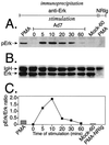
Kinetics of phosphorylation of Erk by Ad7. Serum-starved A549 cells were exposed to Ad7 at an MOI of 50 for various times prior to preparation of cellular lysates and Erk1/2 immunoprecipitation. PMA-exposed cells (60 min), mock-infected cells (either time zero or 60 min), and cells exposed to PMA but immunoprecipitated with NRIg (PMA-NRIg) were used as positive and negative controls. (A) Western blot performed with antibody specific for dually phosphorylated Erk1/2. (B) Same blot stripped and reprobed with pan-anti-Erk1/2 antibody. (C) Activation of Erk as determined by ratio of phospho-Erk to total Erk for each immunoprecipitate. In all cases, bands for both p44 and p42 Erks were included in the calculations. The data shown are representative of three separate experiments.
FIG. 3
FIG. 3
Inhibition of Ad induction of Erk by MEK inhibitor. Serum-starved A549 cells preincubated with various doses of the MEK inhibitor PD98059 were exposed to Ad7 at an MOI of 50. PMA-stimulated or mock-infected cells preincubated with or without PD98059 were used as positive and negative controls. Cells exposed to PMA immunoprecipitated with NRIg (PMA-NRIg) were used as a control for immunoprecipitation. (A) Immunoblot of mock-infected, Ad7-infected, or PMA-stimulated cell extracts probed with anti-phospho-Erk1/2 antibody. (B) Same blot as in panel A stripped and reprobed with pan-anti-Erk1/2 antibody. (C) Ratio of pErk to Erk, determined as for Fig. 2. The data are representative of four separate experiments.
FIG. 4
FIG. 4
Ad7 induces Erk activity. A549 cells were mock infected, PMA stimulated, or Ad7 infected (MOI = 50) in the presence or absence of 25 μM PD98059 for 20 min prior to the preparation of lysates. The lysates were immunoprecipitated overnight with anti-Erk1/2 antibody or with rabbit IgG NRIg. Immunoprecipitated Erk1/2 was then analyzed for activity by an in vitro kinase assay as described in Materials and Methods. (A) Phosphorylated MBP. (B) Plot of Storm PhosphorImager units for each sample analyzed. +, present; −, absent.
FIG. 5
FIG. 5
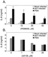
Inhibition of Ad induction of IL-8 protein by MEK inhibitor. (A) Serum-starved A549 cells preincubated with increasing doses of the MEK inhibitor PD98059 were exposed to Ad7 at an MOI of 50 or to virus-free buffer in medium plus 0.5% FBS for 8 h prior to measurement of supernatant IL-8 by ELISA in triplicate (see Materials and Methods). Cells exposed to 100 ng of PMA/ml were used as a positive control. The data are expressed as the mean + standard error of the mean of four separate experiments. (B) A549 cells were incubated as described above with the indicated amounts of U0126 and stimulated with Ad7 or PMA (100 ng/ml) for 4 h. IL-8 was measured in triplicate in supernatants by ELISA and is given as nanograms per milliliter of culture supernatant.
FIG. 6
FIG. 6
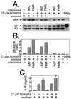
PD98059 inhibition is reversible. Serum-starved A549 cells were preincubated with 50 μM PD98059 or diluent for 30 min and then washed with PBS. The cells were exposed to Ad7 at an MOI of 50 or to virus-free buffer for 20 min prior to the measurement of Erk activation or for 24 h prior to the measurement of IL-8 release (see Materials and Methods). Cells exposed to 100 ng of PMA/ml and cells exposed to PMA immunoprecipitated with NRIg were used as positive and negative controls, respectively. (A) Immunoblot of control, Ad7-infected, or PMA-stimulated and washed cell extracts stained with anti-phospho-Erk1/2 antibody (top) and same membrane stripped and reprobed with anti-Erk1/2 antibody (bottom). The Erk experiment included cells stimulated in the presence of PD98059 after being washed. (B) Activation of Erk as determined by ratio of phospho-Erk to total Erk for each immunoprecipitate. (C) IL-8 levels in supernatants of cells treated as above. The data are the mean + standard error of the mean of four separate experiments. +, present; −, absent.
FIG. 7
FIG. 7
Activation of IL-8 promoter activity by Ad7 is dependent upon activation of Ras pathway. A549 cells were transfected with the Gene Porter 2 system with either IL-8Luc alone or in combination with N17Ras in fourfold excess. Cells were mock or Ad7 infected for 6 h, and luciferase activity was measured. One set of cells transfected with −162 to +44 IL-8Luc alone was preincubated with the MEK inhibitor U0126 (25 μM) for 30 min prior to mock or Ad7 infection.
FIG. 8
FIG. 8
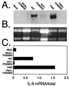
Induction of endogenous IL-8 mRNA levels by Ad7 is inhibited by PD98059. A549 cells were exposed to Ad7 at an MOI of 50 for 2 h in the presence or absence of 25 μM PD98059. PMA (100 ng/ml) was used as a positive control. RNA was extracted and prepared for Northern analysis as described in Materials and Methods. (A) RNA blot probed for IL-8 mRNA. (B) RNA gel of the blot in panel A stained with ethidium bromide. (C) IL-8 mRNA levels expressed as a ratio of IL-8 mRNA to rRNA visualized for each condition.

References

    1. Abbandanzo S L, English C K, Kagan E, McPherson R A. Fatal adenovirus pneumonia in a newborn identified by electron microscopy and in situ hybridization. Arch Pathol Lab Med. 1989;113:1349–1353. - PubMed
    1. Albanyan E A, Vallejo J G, Smith C W, Edwards M S. Nonopsonic binding of type III group B streptococci to human neutrophils induces interleukin-8 release mediated by the p38 mitogen-activated protein kinase pathway. Infect Immun. 2000;68:2053–2060. - PMC - PubMed
    1. Alessi D R, Cuenda A, Cohen P, Dudley D T, Saltiel A R. PD 098059 is a specific inhibitor of the activation of mitogen-activated protein kinase kinase in vitro and in vivo. J Biol Chem. 1995;270:27489–27494. - PubMed
    1. Arnold R, Humbert B, Werchau H, Gallati H, Konig W. Interleukin-8, interleukin-6, and soluble tumour necrosis factor receptor type I release from a human pulmonary epithelial cell line (A549) exposed to respiratory syncytial virus. Immunology. 1994;82:126–133. - PMC - PubMed
    1. Baggiolini M, Dewal B, Moser B. Interleukin-8 and related chemotactic cytokines—CXC and CC chemokines. Adv Immunol. 1994;33:97–179. - PubMed

Publication types

MeSH terms