. 2001 Jul;75(14):6676-81.
doi: 10.1128/JVI.75.14.6676-6681.2001.

Viral replicase gene products suffice for coronavirus discontinuous transcription

Affiliations

Viral replicase gene products suffice for coronavirus discontinuous transcription

V Thiel et al. J Virol. 2001 Jul.

Abstract

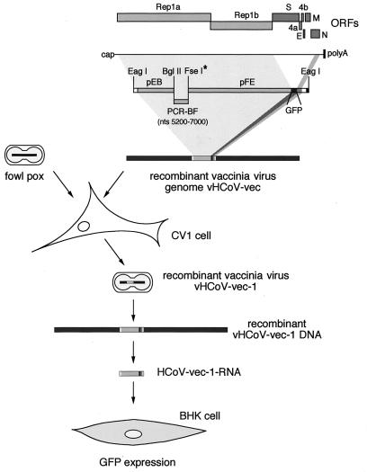
We have used vaccinia virus as a vector to clone a 22.5-kbp cDNA that represents the 5' and 3' ends of the human coronavirus 229E (HCoV 229E) genome, the HCoV 229E replicase gene, and a single reporter gene (coding for green fluorescent protein [GFP]) located downstream of a regulatory element for coronavirus mRNA transcription. When RNA transcribed from this cDNA was transfected into BHK-21 cells, a small percentage of cells displayed strong fluorescence. A region of the mRNA encoding GFP was amplified by PCR and shown to have the unique mRNA leader-body junction indicative of coronavirus-mediated transcription. These data show that the coronavirus replicase gene products suffice for discontinuous subgenomic mRNA transcription.


Figures

FIG. 1
FIG. 1
Strategy for the construction of a coronavirus-based vector RNA that mediates the expression of GFP. The structural relationship among the HCoV 229E ORFs, HCoV 229E genomic RNA, plasmid, PCR cDNA fragments, and in vitro RNA transcripts is shown. The position of three silent mutations, which create a unique FseI (∗) site, is depicted in the recombinant cDNA fragments. Relevant restriction sites are indicated. The cDNA fragments pEB, PCR-BF (which represents the HCoV 229E genomic region [nt 5200 to 7000] that is unstable in bacterial cloning systems), and pFE were assembled by in vitro ligation with the restriction sites BglII and FseI. Subsequent ligation of the resulting cDNA with NotI-cleaved vNotI/tk vaccinia virus DNA produced the recombinant vaccinia virus vHCoV-vec DNA. Recombinant vaccina virus vHCoV-vec-1 was recovered and RNA transcripts were produced in vitro by using genomic vHCoV-vec-1 DNA and bacteriophage T7 RNA polymerase. The HCoV-vec-1 RNA was transfected into BHK-21 cells and monitored for the expression of GFP (see text for details).
FIG. 2
FIG. 2
Cloning of HCoV-vec cDNA in the vaccinia virus genome. (a) Assembly of a 22.5-kbp HCoV-vec cDNA by in vitro ligation. DNA fragments EF and FE were ligated and analyzed by PFGE in comparison with a high-molecular-weight DNA marker (Life Technologies, Karlsruhe, Germany). The ligation substrates and products are illustrated. The relevant restriction sites and the sizes of DNA fragments are shown. AP indicates treatment of DNA fragment ends with alkaline phosphatase. (b) Forced ligation of recombinant vaccinia virus genomes. PFGE analysis of the ligation reaction containing EagI-cleaved, dephosphorylated HCoV-vec insert cDNA and NotI-cleaved vaccinia virus vNotI/tk DNA in the presence of NotI enzyme is shown. The accumulated ligation products, comprised of two vaccinia virus DNA arms and a copy of the HCoV-vec insert cDNA (long/insert/long, long/insert/short, or short/insert/short), as well as relevant substrates and intermediate ligation products, are indicated. DNA bands that are not indicated represent substrates and predicted minor ligation products comprised of pEB, PCR-BF, pFE, and vNotI/tk DNA fragments. (c) Southern blot analysis of 10 random vaccinia virus vHCoV-vec clones. DNA from CV-1 cells infected with vNotI/tk (lane vNot) or recombinant vHCoV-vec clones was digested with HindIII and analyzed by Southern blotting with a random-primed 32P DNA probe representing the HCoV 229E genome between nt 1048 and 20582. Lane M shows a HindIII-digested DNA fragment generated by RT-PCR from HCoV 229E poly(A)-containing RNA. This 19.5-kbp cDNA fragment represents the HCoV genome between nt 1048 and 20582. The two patterns of hybridization (cf. lanes 1 to 6 and 9 with lanes 7, 8, and 10) represent the two possible insert orientations.
FIG. 3
FIG. 3
Expression of GFP by using HCoV-vec-1 RNA (a) The structural relationship among HCoV-vec-1 ORFs, the in vitro-transcribed HCoV-vec-1 RNA, and the intracellular mRNA produced by coronavirus replicase-mediated discontinuous transcription is illustrated together with the predicted intracellular translation products (i.e., the HCoV 229E replicase and GFP). Additionally, 1 μg of capped RNA transcribed from vHCoV-vec-1 DNA in vitro was visualized by ethidium bromide staining after agarose gel electrophoresis. (b) GFP expression analyzed by fluorescence microscopy of BHK-21 cells transfected with HCoV-vec-1 RNA. (c) The TAS region of the intracellular mRNA was sequenced by RT-PCR amplification and cycle sequencing. The sequence corresponding to HCoV 229E leader (L), the TAS region, and the first 10 nt of the GFP-ORF is shown.

References

    1. Agapov E V, Frolov I, Lindenbach B D, Pragai B M, Schlesinger S, Rice C M. Noncytopathic Sindbis virus RNA vectors for heterologous gene expression. Proc Natl Acad Sci USA. 1998;95:12989–12994. - PMC - PubMed
    1. Almázan F, González J M, Pénzes Z, Izeta A, Calvo E, Plana-Durán J, Enjuanes L. Engineering the largest RNA virus genome as an infectious bacterial artificial chromosome. Proc Natl Acad Sci USA. 2000;97:5516–5521. - PMC - PubMed
    1. Ausubel F, Brent R, Kingston R E, Moore D D, Seidman J G, Smith J A, Struhl K. Current protocols in molecular biology. New York, N.Y: John Wiley & Sons; 1987.
    1. Baric R S, Nelson G W, Fleming J O, Deans R J, Keck J G, Casteel N, Stohlman S A. Interactions between coronavirus nucleocapsid protein and viral RNAs: implications for viral transcription. J Virol. 1988;62:4280–4287. - PMC - PubMed
    1. de Vries A F, Horzinek M C, Rottier P J M, de Groot R J. The genome organization of the Nidovirales: similarities and differences between arteri-, toro-, and coronaviruses. Semin Virol. 1997;8:33–47. - PMC - PubMed

Publication types

LinkOut - more resources