. 2001 Jul;21(14):4528-43.
doi: 10.1128/MCB.21.14.4528-4543.2001.

Site-specific recognition of a 70-base-pair element containing d(GA)(n) repeats mediates bithoraxoid polycomb group response element-dependent silencing

Affiliations

Site-specific recognition of a 70-base-pair element containing d(GA)(n) repeats mediates bithoraxoid polycomb group response element-dependent silencing

J W Hodgson et al. Mol Cell Biol. 2001 Jul.

Abstract

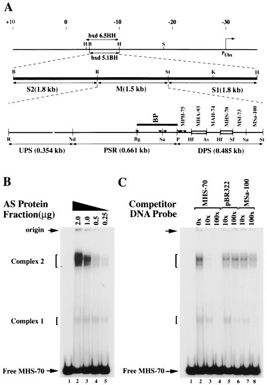
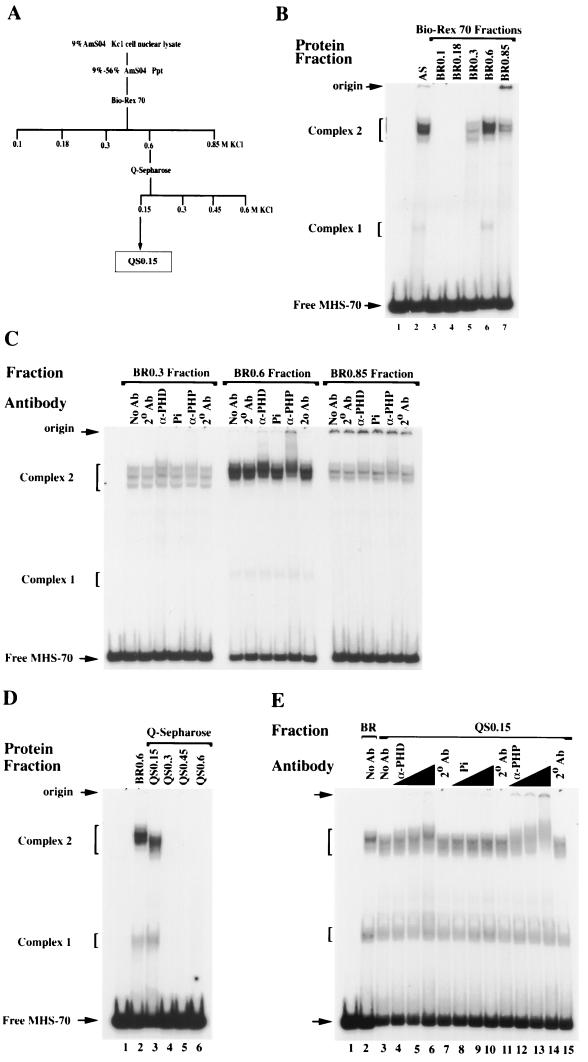
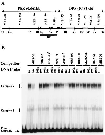
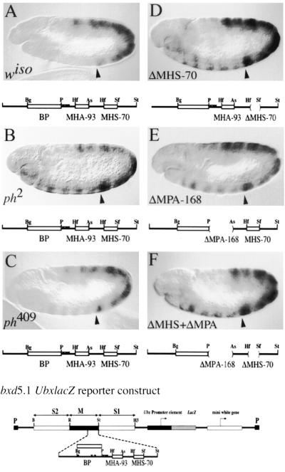
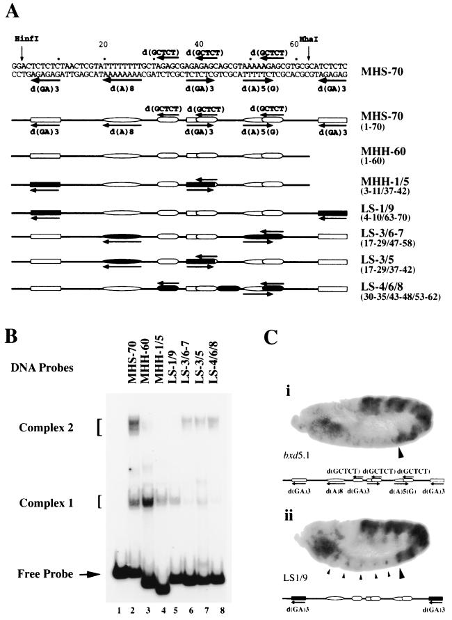
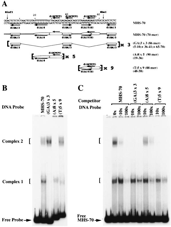
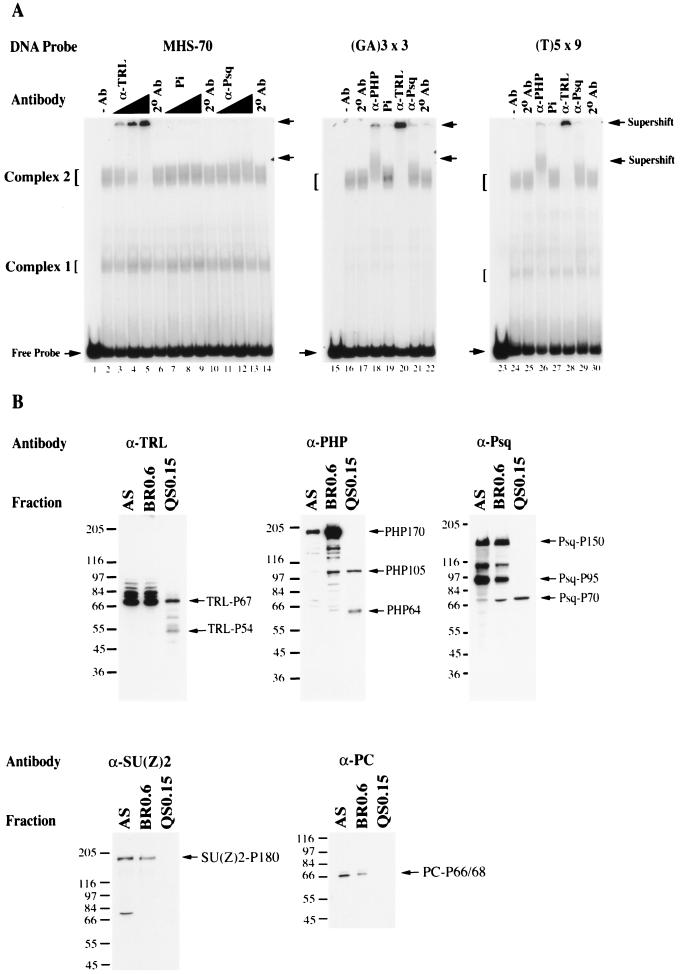
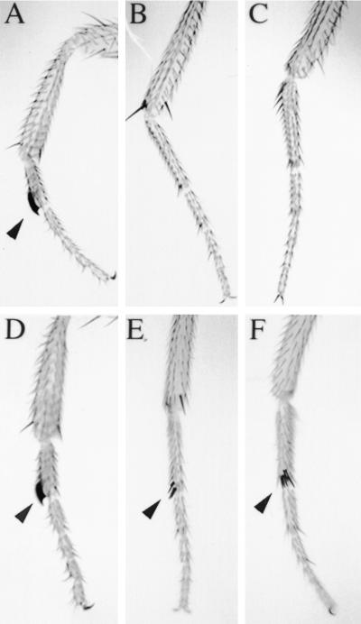
Polycomb group proteins act through Polycomb group response elements (PREs) to maintain silencing at homeotic loci. The minimal 1.5-kb bithoraxoid (bxd) PRE contains a region required for pairing-sensitive repression and flanking regions required for maintenance of embryonic silencing. Little is known about the identity of specific sequences necessary for function of the flanking regions. Using gel mobility shift analysis, we identify DNA binding activities that interact specifically with a multipartite 70-bp fragment (MHS-70) downstream of the pairing-sensitive sequence. Deletion of MHS-70 in the context of a 5.1-kb bxd Polycomb group response element derepresses maintenance of silencing in embryos. A partially purified binding activity requires multiple, nonoverlapping d(GA)(3) repeats for MHS-70 binding in vitro. Mutation of d(GA)(3) repeats within MHS-70 in the context of the 5.1-kb bxd PRE destabilizes maintenance of silencing in a subset of cells in vivo but gives weaker derepression than deletion of MHS-70. These results suggest that d(GA)(3) repeats are important for silencing but that other sequences within MHS-70 also contribute to silencing. Antibody supershift assays and Western analyses show that distinct isoforms of Polyhomeotic and two proteins that recognize d(GA)(3) repeats, the TRL/GAGA factor and Pipsqueak (Psq), are present in the MHS-70 binding activity. Mutations in Trl and psq enhance homeotic phenotypes of ph, indicating that TRL/GAGA factor and Psq are enhancers of Polycomb which have sequence-specific DNA binding activity. These studies demonstrate that site-specific recognition of the bxd PRE by d(GA)(n) repeat binding activities mediates PcG-dependent silencing.


Figures

FIG. 1
FIG. 1
Identification of binding activities recognizing MHS-70. (A) Schematic representation of the organization of the bxd PRE. The start site of Ubx transcription is marked by a bent arrow, the coordinates of Bender et al. (2) are marked at the top, and the positions of bxd6.5HH and bxd5.1BH are indicated. The functional regions of bxd5.1, S1, M, and S2 are indicated on the second line and are described in the text. A detailed restriction map of the M element is shown on the third line. Fragments are named by the first letters of the restriction enzymes defining them, followed by their length in base pairs. PSR corresponds to the pairing-sensitive element defined by Sigrist and Pirrotta (52), and the dark line marked BP identifies the fragment described by Horard et al. (26). Small triangles mark the location of PHO binding sites (20). Open boxes represent binding sites identified in fragment DPS. Restriction site abbreviations: As, AseI; B, BamHI; Bg, BglI; H, HindIII; Hf, HinfI; K, KpnI; Nd, NdeI; P, PstI; R, EcoRI; S, SalI; Sa, Sau3A; Sf, SfaNI; St, StyI. (B) Gel mobility shift analyses of complexes formed at MHS-70 with no protein (lane 1) or with decreasing amounts of the AS fraction of KcI cell nuclear extract (lanes 2 to 5). Positions of nucleoprotein complexes formed are shown on the left. (C) Competitive gel mobility shift analysis of complexes formed at MHS-70 (with 0.6 μg of the AS fraction) in the presence of a molar excess of unlabeled, nonspecific (pBR322 BamHI-SalI 276-bp fragment) and specific (MSa-100) competitor DNA fragment (shown on top). Complex 2 formation is sequence specific.
FIG. 2
FIG. 2
Fractionation and supershift analysis of MHS-70 binding activities. (A) Chromatographic fractionation scheme for MHS-70 binding activities. (B) Gel mobility shift analysis of MHS-70 binding activities present in Bio-Rex 70 fractions, eluted stepwise with buffers containing 0.1 to 0.85 M KCl. Lanes 1 and 2, no protein and 0.6 μg of AS fraction, respectively; lane 6, 0.3 μg of BR0.6; lanes 3, 4, 5, and 7, 1.6 μg of the Bio-Rex 70 fractions shown above the gel. Low mobility complexes migrating close to the origin are detected with fractions eluted at 0.85 M KCl. (C) Supershift analysis of BR0.3, BR0.6, and BR0.85. No antibody (No Ab), antibody for PHD (α-PHD), preimmune serum (Pi), and antibody specific for PHP (α-PHP) at a titer of 1:37.5 were incubated in the binding reactions of the Bio-Rex 70 fractions to MHS-70 in the presence of secondary antibody (2° Ab), and changes in mobility were monitored in the gel shift assay. Partial supershifts of complex 2 are clearly detected in BR 0.6 and are not detected in BR0.3, and are weakly detected in BR0.85. (D) Mobility shift analysis of QS0.15, QS0.3, QS0.45, and QS0.6 with MHS-70. Lanes 1 and 2, no protein and 0.3 μg of BR0.6 respectively; lanes 3 to 6, 0.25 μg of protein from each Q Sepharose fraction. The MHS-70 binding activity is only detectable in QS0.15. (E) Supershift analysis of the QS0.15 binding activity (0.25 μg of protein) with anti-PH antibodies (lanes 4 to 6 and 12 to 14) at titers of 1:75 (lanes 4 and 12), 1:37.5 (lanes 5 and 13), and 1:10 (lanes 6 and 14). Both anti-PH antibodies but not the equivalent titer of preimmune serum (lanes 8 to 10) cause significant supershifts of complex 2.
FIG. 3
FIG. 3
Competitive gel mobility shift analysis of the QS0.15 activity with M element fragments. (A) DNA fragments from the bxd M element, indicated on the top line, were used as competitors in the binding reaction of 0.25 μg of QS0.15 with MHS-70. Abbreviations for restriction enzymes and nomenclature of fragments are given in the legend to Fig. 1A, with the exception of Bf (BfaI). Fragments from the PSR are indicated above the line. (B) Competitive gel mobility shift analysis of complexes formed at MHS-70 in the presence of a molar excess of unlabeled fragments from the bxd M element. As expected, MHS-70 completely inhibits the formation of complex 2. Partial inhibition of complex 2 formation is detected with fragments MHA-93 and MSP-61 (marked with asterisks).
FIG. 4
FIG. 4
Mutational analysis of bxd5.1 UbxlacZ expression in germband-extended embryos. Germband-extended embryos are mounted anterior to the left, dorsal side up. Expression of LacZ was detected immunohistochemically. The anterior boundary of PS6 is marked with an arrowhead. The wild-type bxd5.1 UbxlacZ reporter, consisting of the 5.1-kb bxd PRE, the Ubx promoter, and the lacZ and white reporters flanked by P element long terminal repeats, is shown below the figure, with the DPS fragment expanded to show detail. The DPS fragment is indicated below each panel to show if it is wild type (A to C) or to show the positions and numbers of deletions in mutant constructs (indicated by parentheses in panel D to F). The same bxd5.1 UbxlacZ reporter, which exhibits complete silencing anterior to PS6, is used in panels A to C. (A) Wild-type embryo from a line exhibiting complete silencing of the reporter in PS1 to PS5 and in the head. (B) ph2 mutant embryo, showing partial derepression of bxd5.1 in PS1 to 5 and in the head. (C) ph409 mutant embryo, showing weak derepresion of bxd5.1 in PS 1 to 5. (D) bxd5.1ΔMHS-70 embryo in a wild-type background showing partial derepression of the reporter in PS1 to PS5. (E) bxd5.1ΔMPA-168 embryo exhibiting a derepression pattern similar to that seen in bxd5.1ΔMHS-70. (F) bxd5.1ΔMHS-70 + ΔMPA-168 embryo showing a derepression pattern similar to that seen in either single mutant (compare to panels D and E). Notation is as for Fig. 1A.
FIG. 5
FIG. 5
Gel shift and in vivo analysis of MHS-70 linker-scanning mutations. (A) Schematic representation of MHS-70 and mutated derivatives. Details of mutations are given in the text. The sequences of interest are marked in the top line and schematically represented in the second line. Sites of mutations are indicated by filled regions in the schematic representations of mutated MHS-70 fragments in lines 3 to 7. Coordinates of the substitution sites are shown on the right in parentheses. (B) Mobility shift analysis of MHS-70 mutants using 0.25 μg of QS0.15. The identities of mutant fragments used in each lane are shown in panel A. Complex 2 formation is abolished by combined substitutions of the d(GA)3 repeats. (C) Derepression caused by d(GA)3 substitution mutations of MHS-70 in bxd5.1 PRE. Conditions and reporters are explained in the legend to Fig. 4. The anterior boundary of PS6 is marked with a large arrowhead. The MHS-70 fragment is schematically represented below each panel. (i) bxd5.1 UbxlacZ from a line exhibiting spotty misexpression in PS1 to PS5 and partial derepression in the head. The PS6 boundary is maintained. (ii) bxd5.1 UbxlacZ mutant for two d(GA)n sites of MHS-70, LS-1/9 (illustrated in Fig. 5A). Note the clear derepression in PS1 to PS5 in a distinct subset of cells (small arrowheads), plus increased derepression in the head.
FIG. 6
FIG. 6
Gel shift analysis of MHS-70 MSR elements. (A) Schematic representation of oligomers containing MSR elements used in binding studies. Lines 1 and 2 are the same as in Fig. 5A; lines 3 to 5 show the sequences used to construct oligonucleotides. Sequences of the oligonucleotides are given in Table 1. (B) Mobility shift analysis of oligonucleotides derived from MHS-70 with 0.25 μg of QS0.15. Identities of the oligonucleotides are shown in panel A, and coordinates of the repeat elements are shown on the right in parentheses. Complex 2 is formed at (GA)3 × 3 and (T)5 × 9, and complex 1 is formed at (A)8 × 5 and (T)5 × 9. (C) Competitive mobility shift assay. Molar excesses of the oligonucleotides shown in panel A were used as competitors for the binding of 0.25 μg of QS0.15 to MHS-70. (GA) 3 × 3 and (T)5 × 9 abolished the formation of complex 2. The (A)8 × 5 oligomer did not inhibit the formation of complex 2.
FIG. 7
FIG. 7
Supershift and Western analysis of QS0.15. (A) Secondary antibody (2° Ab) in the absence or presence of primary antibodies to PH (α-PHP), TRL/GAF (α-TRL), or Psq (α-Psq) or preimmune serum (Pi) was incubated in binding reactions of 0.25 μg of QS0.15 with MHS-70, (GA)3 × 3, or (T)5 × 9, and the nucleoprotein complexes were examined in a mobility shift assay. Lanes 1, 15, and 23 no protein; lanes 2, 16, and 24, no antibody (−Ab); lanes 6, 10, 14, 17, 22, 25, and 30, only secondary antibody (2° Ab). In the MHS-70 binding reactions, α-TRL titers were 1:240 (lanes 3), 1:120 (lane 4), and 1:60 (lane 5); Pi and α-Psq titers were 1:30 (lanes 7, and 11), 1:10 (lanes 8 and 12), and 1:7.5 (lanes 9 and 13). In the (GA)3 × 3 or (T)5 × 9 binding reactions, α-PHP was used at a titer of 1:10 (lanes 18 and 26), α-TRL was used at 1:60 (lanes 20 and 28), and α-Psq or Pi was used at 1:7.5 (lanes 21 and 29 or lanes 19 and 27). Antibodies to TRL and PHP caused a strong supershift in reactions of all fragments tested, and anti-Psq caused a detectable, partial supershift in reactions of the same fragments. (B) Western analysis of 20 μg of protein derived from AS, BR0.6, and QS0.15 fractions described in Fig. 2A. The fractions were separated by SDS-PAGE on a 9% gel, transferred to nitrocellulose, and probed with the antibodies indicated in panel A as described in Materials and Methods. Specific isoforms of each protein were enriched for and detected in QS0.15 are shown on the right of each gel; positions of protein molecular mass markers (in kilodaltons) (Sigma) are shown on the left.
FIG. 8
FIG. 8
Trl and psq mutations enhance the extra sex combs phenotype of ph mutants. Wild-type Drosophila males have two sex combs (marked by arrowheads): one on each first leg (prothoracic leg) (A) and absent on the second (mesothoracic) (B) and third (metathoracic) (C) legs. Males hemizygous for ph mutations exhibit posterior homeotic transformations where the second and third legs are frequently transformed into the first, thus increasing the total number of sex combs per male fly. Male flies transheterozygous for trl or psq and ph mutations exhibit an enhancement of the extra sex combs phenotype of ph. For example, ph2/Y; Trl62/+ males always exhibit sex combs on the first legs (D), frequently on the second legs (E), and sometimes on the third legs (F).

References

    1. Akam M. The molecular basis for metameric pattern in the Drosophila embryo. Development. 1987;101:1–22. - PubMed
    1. Bender W, Akam M, Karch F, Beachy P A, Peifer M, Spierer P, Lewis E B, Hogness D S. Molecular genetics of the Bithorax complex in Drosophila melanogaster. Science. 1983;221:23–29. - PubMed
    1. Benyajati C, Mueller L, Xu N, Pappano M, Gao J, Mosammaparast M, Conklin D, Granok H, Craig C, Elgin S. Multiple isoforms of GAGA factor, a critical component of chromatin structure. Nucleic Acids Res. 1997;25:3345–3353. - PMC - PubMed
    1. Bhat K M, Farkas G, Karch F, Gyurkovics H, Gausz J, Schedl P. The GAGA factor is required in the early Drosophila embryo not only for transcriptional regulation but also for nuclear division. Development. 1996;122:1513–1522. - PubMed
    1. Biggin M D, Tjian R. Transcription factors that activate the Ultrabithorax promotor in developmentally staged extracts. Cell. 1988;53:699–711. - PubMed

Publication types

LinkOut - more resources