. 2001 Jul;21(14):4829-36.
doi: 10.1128/MCB.21.14.4829-4836.2001.

Proteinuria and perinatal lethality in mice lacking NEPH1, a novel protein with homology to NEPHRIN

Affiliations

Proteinuria and perinatal lethality in mice lacking NEPH1, a novel protein with homology to NEPHRIN

D B Donoviel et al. Mol Cell Biol. 2001 Jul.

Abstract

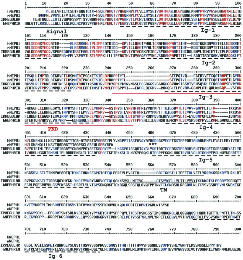
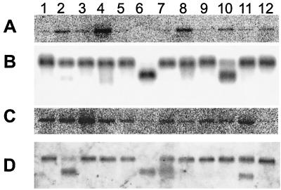
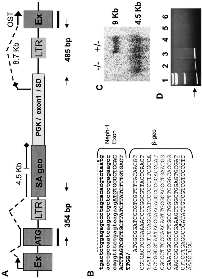
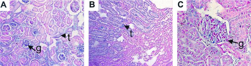
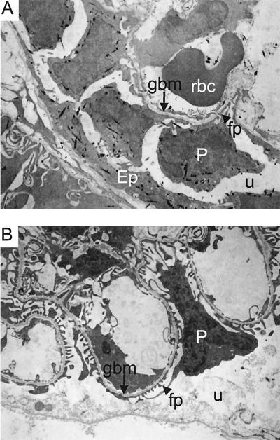
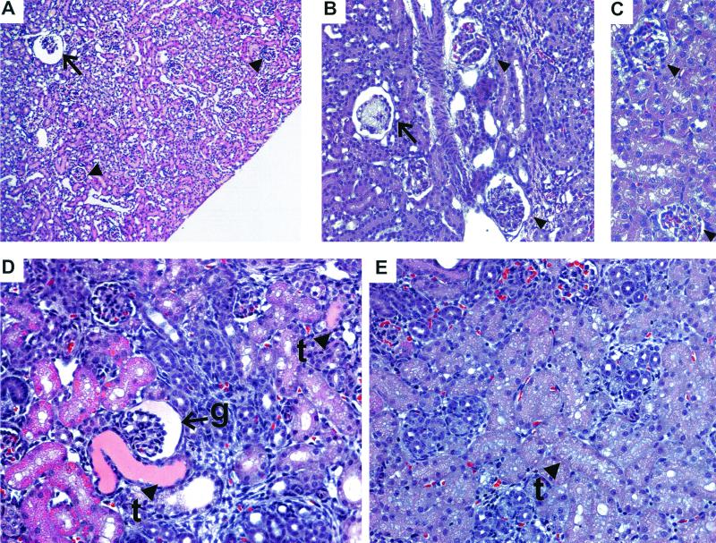
A high-throughput, retrovirus-mediated mutagenesis method based on gene trapping in embryonic stem cells was used to identify a novel mouse gene. The human ortholog encodes a transmembrane protein containing five extracellular immunoglobulin-like domains that is structurally related to human NEPHRIN, a protein associated with congenital nephrotic syndrome. Northern analysis revealed wide expression in humans and mice, with highest expression in kidney. Based on similarity to NEPHRIN and abundant expression in kidney, this protein was designated NEPH1 and embryonic stem cells containing the retroviral insertion in the Neph1 locus were used to generate mutant mice. Analysis of kidney RNA from Neph1(-/-) mice showed that the retroviral insertion disrupted expression of Neph1 transcripts. Neph1(-/-) pups were represented at the expected normal Mendelian ratios at 1 to 3 days of age but at only 10% of the expected frequency at 10 to 12 days after birth, suggesting an early postnatal lethality. The Neph1(-/-) animals that survived beyond the first week of life were sickly and small but without edema, and all died between 3 and 8 weeks of age. Proteinuria ranging from 300 to 2,000 mg/dl was present in all Neph1(-/-) mice. Electron microscopy demonstrated NEPH1 expression in glomerular podocytes and revealed effacement of podocyte foot processes in Neph1(-/-) mice. These findings suggest that NEPH1, like NEPHRIN, may play an important role in maintaining the structure of the filtration barrier that prevents proteins from freely entering the glomerular urinary space.


Figures

FIG. 1
FIG. 1
Alignment of NEPH1 with homologous proteins. The human NEPH1 (hNEPH1; GenBank accession no. AYO17369), a truncated mouse NEPH1 (mNEPH1; GenBank accession no. AYO17368), the D. melanogaster irregular chiasmata (IRREGULAR; GenBank accession no. L11040), and the human NEPHRIN (hNEPHRIN; GenBank accession no. AF190637) proteins were aligned using the MultiAlin version 5.4.1 program (http://www.toulouse.inra.ft/multalin). The signal and transmembrane (TM) domains in the hNEPH1, mNEPH1, and IRREGULAR proteins, identified by DNAStar Protean V. 4.0, are underlined. Ig-like domains (Ig) are marked by broken lines, and the PKD domain is doubly underlined; these domains were identified using the Pfam search engine (http://pfam.wustl.edu). The RGD sequence of NEPH1 is marked by asterisks. The hNEPHRIN protein sequence extends an additional 459 amino acid residues beyond what is shown here.
FIG. 2
FIG. 2
Neph1 expression in human and mouse tissues. (A) Northern blot showing a 9-kb Neph1 transcript in adult human brain (lane 1), colon (lane 2), heart (lane 3), kidney (lane 4), liver (lane 5), lung (lane 6), muscle (lane 7), placenta (lane 8), small intestine (lane 9), spleen (lane 10), stomach (lane 11), and testis (lane 12). (B) The same blot as in panel A hybridized with a probe to the human β-actin gene for RNA loading control. (C) Northern blot showing a 9-kb Neph1 transcript in adult mouse brain (lane 1), heart (lane 2), kidney (lane 3), liver (lane 4), lung (lane 5), muscle (lane 6), skin (lane 7), small intestine (lane 8), spleen (lane 9), stomach (lane 10), testis (lane 11), and thymus (lane 12). (D) The same blot as in panel C hybridized with a human β-actin probe.
FIG. 3
FIG. 3
Retroviral insertion into the Neph1 locus. (A) Structure of the gene trap vector integration site in the Neph1 locus. The retroviral construct, which contains flanking viral long terminal repeats (LTRs), is inserted in the intron downstream of the ATG-containing Neph1 exon. Transcripts initiating at the Neph1 promoter (indicated by an arrow followed by broken and solid lines) encode a 4.5-kb message that utilizes the splice acceptor (SA) site upstream of the β-geo fusion gene. Transcripts initiating from the PGK promoter utilize the splice donor site in the inserted heterologous exon 1-splice donor (SD)-intron 1 cassette and splice into the downstream Neph1 exon (EX) to produce a roughly 8.7-kb RNA which is detectable only in ES cells and is absent in vivo (see text). The heterologous exon 1 is a noncoding exon that contains a natural in-frame stop codon (black diamond) that eliminates the likelihood of expression of a protein from the trapped Neph1 locus. A probe spanning the ATG-containing exons and downstream exons of Neph1 (black bars) was used to perform the Northern blot shown in Fig. 2 and 3C. The OST sequence is represented by a double line. The primers used to clone the Neph1–β-geo and the exon 1-Neph1 fusion transcript splice junctions are indicated by arrows and are expected to produce 354- and 485-bp bands, respectively; the sequence of the 5′ transcript is given in panel B. (B) Sequence of the hybrid transcript spanning the splice junction of the Neph1 ATG-containing exon and the β-geo-coding region. The ATG in the Neph1 coding region (boldface) is underlined, and the primers used for RT-PCR of this fragment are designated by broken arrows. (C) Northern blot of kidney RNA obtained from 1-day-old mice bearing either one copy (+/−) or two copies (−/−) of the trapped Neph1 locus. The blot, hybridized with a probe spanning the Neph1 ATG-containing and downstream exons (see panel A), shows RNA transcripts of 9 and 4.5 kb. (D) Ethidium bromide-stained gel showing RT-PCR products from the heterologous exon 1-Neph1 fusion transcripts. Lane 1 contains molecular-weight standards. The expected band size of 485 bp (see panel A) is clearly observed in the RNA samples from the ES cell line with the Neph1 mutation (lanes 2 and 3). This band is undetectable in kidney RNA from Neph1+/+ and Neph1+/− mice (lanes 4 and 6, respectively) and is negligible in kidney RNA from Neph1−/− mice (lane 5).
FIG. 4
FIG. 4
Neph1 expression as indicated by LacZ staining (blue) of kidneys from newborn (A) and adult (B and C) heterozygous mice. (A) Staining in the glomeruli (g, arrow) and proximal tubules (t, arrowhead) of a 1-day-old newborn pup. (B) Staining in the proximal tubules (t, arrowhead) of an adult kidney. (C) Staining in a juxtamedullary glomerulus (g, arrow) of an adult mouse kidney.
FIG. 5
FIG. 5
Electron micrographs of LacZ-stained Neph1−/− kidney. (A) Electron micrograph depicting LacZ stain crystals (black specks) indicating expression of Neph1–β-geo fusion transcripts in the cytoplasm of podocytes (P) and parietal epithelium (Ep) of a Neph1−/− newborn mouse. The glomerular basement membrane (gbm) is marked with an arrow, and a podocyte foot process (fp) is depicted by an arrowhead. There is significant effacement of the podocyte foot processes in Neph1−/− mutant animals (see text and compare to panel B). The urinary space (u) and a red blood cell (rbc) are also noted. (B) Electron micrograph depicting no LacZ stain crystals in a Neph1+/+ kidney. The podocyte (P), glomerular basement membrane (gbm), foot processes (fp), and urinary space (u) are marked as in panel A.
FIG. 6
FIG. 6
Kidney abnormalities in Neph1−/− mice. (A) Dilated (cystic) juxtamedullary glomerulus (arrow) in a 3-week-old Neph1−/− mouse. Cortical glomeruli (arrowheads) look normal. (B) Mesangiolysis (arrow) and mesangial hypercellularity and increased matrix (arrowheads) in glomeruli from 3-week-old Neph1−/− kidney. (C) Normal glomeruli (arrowheads) from a control 3-week-old littermate. (D) Protein-filled tubules (t, arrowheads) and cystic glomerulus (g, arrow) from a newborn Neph1−/− mouse. (E) Control newborn mouse kidney showing normal tubules (t, arrowhead).

References

    1. Anonymous. Guide for the care and use of laboratory animals. Bethesda, Md: National Institutes of Health; 1985.
    1. Bancroft J, Stevens A. Theory and practice of histological techniques. 2nd ed. New York, N.Y: Churchill Livingstone; 1982.
    1. Boute N, Gribouval O, Roselli S, Benessy F, Lee H, Fuchshuber A, Dahan K, Gubler M C, Niaudet P, Antignac C. NPHS2, encoding the glomerular protein podocin, is mutated in autosomal recessive steroid-resistant nephrotic syndrome. Nat Genet. 2000;24:349–354. . (Erratum, 25:125.) - PubMed
    1. Bycroft M, Bateman A, Clarke J, Hamill S J, Sandford R, Thomas R L, Chothia C. The structure of a PKD domain from polycystin-1: implications for polycystic kidney disease. EMBO J. 1999;18:297–305. - PMC - PubMed
    1. Friedrich G, Soriano P. Promoter traps in embryonic stem cells: a genetic screen to identify and mutate developmental genes in mice. Genes Dev. 1991;5:1513–1523. - PubMed

Associated data