. 2001 Jul 3;98(14):8077-82.
doi: 10.1073/pnas.141218998. Epub 2001 Jun 19.

The affective component of pain in rodents: direct evidence for a contribution of the anterior cingulate cortex

Affiliations

The affective component of pain in rodents: direct evidence for a contribution of the anterior cingulate cortex

J P Johansen et al. Proc Natl Acad Sci U S A. .

Abstract

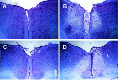
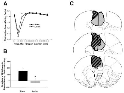
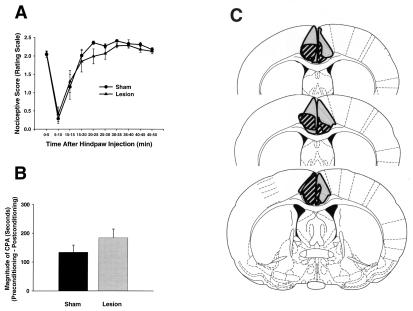
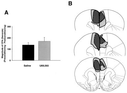
Numerous human and animal studies indirectly implicate neurons in the anterior cingulate cortex (ACC) in the encoding of the affective consequences of nociceptor stimulation. No causal evidence, however, has been put forth linking the ACC specifically to this function. Using a rodent pain assay that combines the hind-paw formalin model with the place-conditioning paradigm, we measured a learned behavior that directly reflects the affective component of pain in the rat (formalin-induced conditioned place avoidance) concomitantly with "acute" formalin-induced nociceptive behaviors (paw lifting, licking, and flinching) that reflect the intensity and localization of the nociceptive stimulus. Destruction of neurons originating from the rostral, but not caudal, ACC reduced formalin-induced conditioned place avoidance without reducing acute pain-related behaviors. These results provide evidence indicating that neurons in the ACC are necessary for the "aversiveness" of nociceptor stimulation.


Figures

Figure 1
Figure 1
Photomicrographs of representative coronal sections through the rostral (A and B) and caudal (C and D) ACC. Sections were stained with cresyl violet. (A) Section from a rostral ACC sham lesion animal as compared with a section taken from a rostral ACC lesion animal (B) at the same antero-posterior level. Sections taken from the same antero-posterior level of caudal ACC sham lesion and caudal ACC lesion animals are compared in C and D, respectively. Clear lesion areas are evidenced by neuronal cell loss and by the proliferation of smaller glial cells (especially apparent around the borders of the lesions).
Figure 2
Figure 2
Formalin-induced pain behaviors and brain lesion maps associated with rats with rostral ACC lesions (n = 8) and rostral ACC sham lesions (n = 10). Data are represented as mean ± SEM. Acute formalin-induced nociceptive scores (rating scale) are shown in A. Magnitude of CPA scores are shown in B. Representations of the largest and smallest lesions in the rostral ACC lesion group are shown in C. Mean percent damage calculations for each hemisphere and a bilateral mean for each experimental group are as follows: left hemisphere, 71% ± 4%; right hemisphere, 51% ± 6%; mean, 62% ± 4%. Rostral ACC lesions completely abolished F-CPA (B) without causing a concomitant reduction in acute formalin-induced nociceptive behaviors (A). (A) *, P < 0.05, Tukey's honestly significant difference test, as compared with rats with sham lesions of the rostral ACC. (B) *, P < 0.05, Student's t test, as compared with rats with sham lesions of the rostral ACC.
Figure 3
Figure 3
Formalin-induced pain behaviors and brain lesion maps associated with rats with caudal ACC lesions (n = 8) and caudal ACC sham lesions (n = 11). Data are represented as mean ± SEM. Acute formalin-induced nociceptive scores are shown in A. Magnitude of CPA scores are shown in B. Representations of the largest and smallest lesions in the caudal ACC lesion group are shown in C. Mean “percent damage” calculations for each hemisphere and a bilateral mean for each experimental group are as follows: left hemisphere, 82% ± 5%; right hemisphere, 67% ± 5%; mean, 74% ± 4%. Caudal ACC lesions had no effect on either acute formalin-induced nociceptive behaviors (A) or F-CPA (B).
Figure 4
Figure 4
CPA produced by a non-nociceptive stimulus (the κ-opioid receptor agonist U69,593) in rats with rostral ACC lesions (n = 9) and rostral ACC sham lesions (n = 8). Data are represented as mean ± SEM. U69,593-induced magnitude of CPA scores are shown in A. Representations of the largest and smallest lesions in the rostral ACC lesion group are shown in B. Mean percent damage calculations for each hemisphere and a bilateral mean for each experimental group are as follows: left hemisphere, 84% ± 4%; right hemisphere, 60% ± 3%; mean, 69% ± 3%. Rostral ACC lesions had no effect on U69,593-induced CPA.

References

    1. Fields H L. Pain. 1999;6,Suppl.:S61–S69. - PubMed
    1. Melzack R, Casey K L. In: The Skin Senses. Kenshalo D, editor. Springfield, IL: Thomas; 1968. pp. 423–439.
    1. Melzack R. Pain. 1975;1:277–299. - PubMed
    1. Price D D. Science. 2000;288:1769–1772. - PubMed
    1. Bushnell M C, Duncan G H, Hofbauer R K, Ha B, Chen J I, Carrier B. Proc Natl Acad Sci USA. 1999;96:7705–7709. - PMC - PubMed

Publication types