Review
. 2001 Jun 19;98(13):7012-7.
doi: 10.1073/pnas.111145498.

Assembly and transport of a premessenger RNP particle

Affiliations
Review

Assembly and transport of a premessenger RNP particle

B Daneholt. Proc Natl Acad Sci U S A. .

Abstract

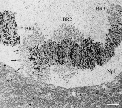
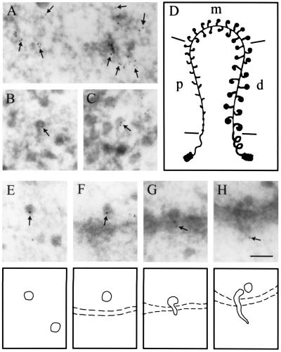
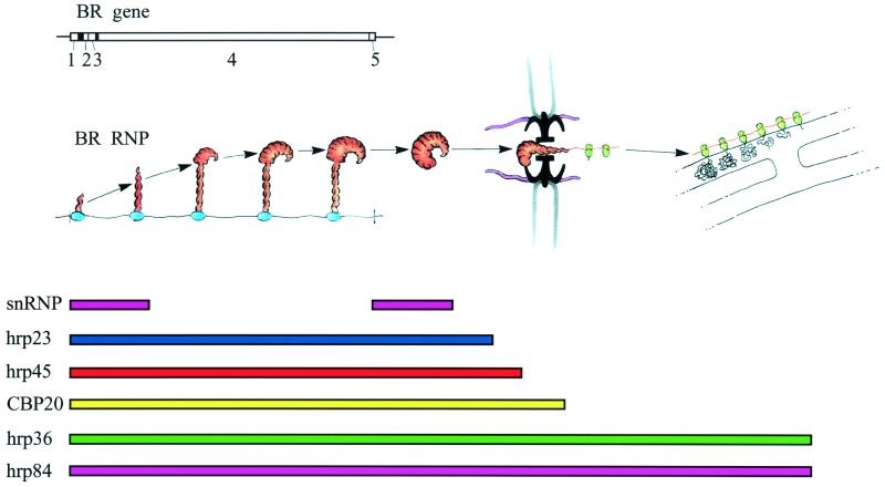
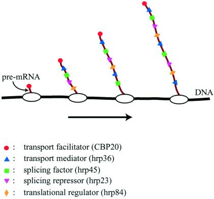
Salivary gland cells in the larvae of the dipteran Chironomus tentans offer unique possibilities to visualize the assembly and nucleocytoplasmic transport of a specific transcription product. Each nucleus harbors four giant polytene chromosomes, whose transcription sites are expanded, or puffed. On chromosome IV, there are two puffs of exceptional size, Balbiani ring (BR) 1 and BR 2. A BR gene is 35-40 kb, contains four short introns, and encodes a 1-MDa salivary polypeptide. The BR transcript is packed with proteins into a ribonucleoprotein (RNP) fibril that is folded into a compact ring-like structure. The completed RNP particle is released into the nucleoplasm and transported to the nuclear pore, where the RNP fibril is gradually unfolded and passes through the pore. On the cytoplasmic side, the exiting extended RNP fibril becomes engaged in protein synthesis and the ensuing polysome is anchored to the endoplasmic reticulum. Several of the BR particle proteins have been characterized, and their fate during the assembly and transport of the BR particle has been elucidated. The proteins studied are all added cotranscriptionally to the pre-mRNA molecule. The various proteins behave differently during RNA transport, and the flow pattern of each protein is related to the particular function of the protein. Because the cotranscriptional assembly of the pre-mRNP particle involves proteins functioning in the nucleus as well as proteins functioning in the cytoplasm, it is concluded that the fate of the mRNA molecule is determined to a considerable extent already at the gene level.


Figures

Figure 1
Figure 1
Electron micrograph showing chromosome IV with its three giant puffs (BRs) in a salivary gland cell from C. tentans. The three BRs (BR1, BR2, and BR3) are indicated as well as the nucleoplasm (Npl) and cytoplasm (Cpl). The arrows mark a few prominent transcription loops (cf. Fig. 2D). (Bar equals 2 μm.)
Figure 2
Figure 2
Intracellular distribution of the cap-binding protein CBP20 in C. tentans salivary gland cells studied by immunoelectron microscopy. The assembly of the BR RNP particle is shown in A–D: proximal portions of the BR gene are displayed in A, distal portions in B and C, and a schematic drawing of the BR gene in D (p, proximal; m, middle; d, distal portions of the gene). The fate of the released BR particles is shown in E–H: BR particles are present in the nucleoplasm (E), at the pore (F), and in an unfolded conformation when passing through the pore (G and H). Gold particles are marked by arrows and indicate the position of CBP20. It should be noted that gold particles are at the leading 5′ end of the BR particle when it passes through the nuclear pore. (Bar equals 100 nm.) Modified from ref. ; produced by permission of The Rockefeller University Press.
Figure 3
Figure 3
Assembly and transport of the BR RNP particle and its relation to a number of BR RNA-associated proteins. The BR particle is assembled on the gene (left), passes through the nucleoplasm, unfolds, and translocates through the nuclear pore (middle). On the cytoplasmic side, the BR RNP fibril becomes engaged in protein synthesis and the polysomes anchor at the endoplasmic reticulum (right). The tripartite nuclear pore complex with its central channel is seen in black and its nuclear and cytoplasmic fibers are presented in pink. The BR gene with its five exons is displayed above the BR particle scheme, and the flow patterns of the BR RNA-associated proteins are outlined below. snRNP, small nuclear RNP. Modified from ref. ; printed with permission from Elsevier Science.
Figure 4
Figure 4
Cotranscriptional loading of proteins onto growing BR pre-mRNA molecules. Some proteins bind to the pre-mRNA with high sequence specificity (e.g., CBP20), whereas others bind with lower specificity along the entire RNA molecule (e.g., the SR protein hrp45 and the 2xRBD-Gly protein hrp36). The RNP fibril formed serves as the substrate for trans-acting factors, and its structure affects a number of mRNA-related processes, including splicing, transport, and translation.

References

    1. Cremer T, Kurz A, Zirbel R, Dietzel S, Rinke B, Schröck E, Speicher M R, Mathieu U, Jauch A, Emmerich P, et al. Cold Spring Harbor Symp Quant Biol. 1973;58:777–792. - PubMed
    1. van Driel R, Wansink D G, van Steensel B, Grande M A, Schul W, de Jong L. Int Rev Cytol. 1995;162A:151–188. - PubMed
    1. Spector D L. Proc Natl Acad Sci USA. 1990;87:147–151. - PMC - PubMed
    1. Zachar Z, Kramer J, Mims I P, Bingham P M. J Cell Biol. 1993;121:729–742. - PMC - PubMed
    1. Fakan S, Puvion E, Spohr G. Exp Cell Res. 1976;99:155–164. - PubMed

Publication types

Substances

LinkOut - more resources