Review
. 2001 Jun 19;98(13):7025-8.
doi: 10.1073/pnas.111145698.

Spatial and temporal control of RNA stability

Affiliations
Review

Spatial and temporal control of RNA stability

A Bashirullah et al. Proc Natl Acad Sci U S A. .

Abstract

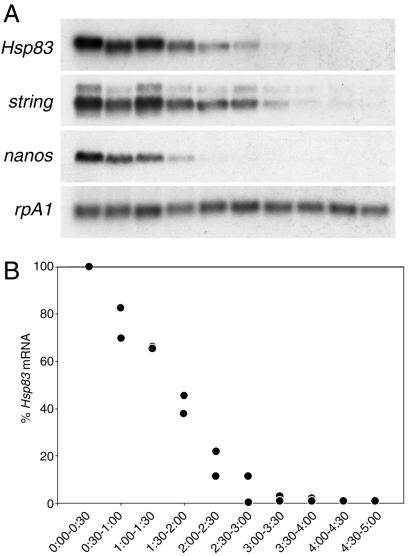
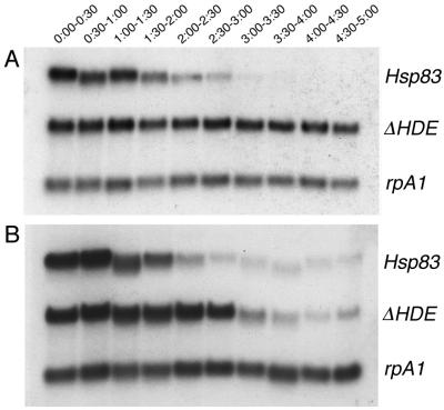
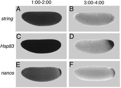
Maternally encoded RNAs and proteins program the early development of all animals. A subset of the maternal transcripts is eliminated from the embryo before the midblastula transition. In certain cases, transcripts are protected from degradation in a subregion of the embryonic cytoplasm, thus resulting in transcript localization. Maternal factors are sufficient for both the degradation and protection components of transcript localization. Cis-acting elements in the RNAs convert transcripts progressively (i) from inherently stable to unstable and (ii) from uniformly degraded to locally protected. Similar mechanisms are likely to act later in development to restrict certain classes of transcripts to particular cell types within somatic cell lineages. Functions of transcript degradation and protection are discussed.


Figures

Figure 1
Figure 1
Time course of maternal transcript degradation in activated, unfertilized eggs. (A) The same Northern blot probed for rpA1 (stable) and string, Hsp83, or nanos (unstable) transcripts. (B) Quantitative analysis of the time course of Hsp83 transcript degradation. The points represent the ratio of Hsp83 transcripts to stable rpA1 transcripts relative to the initial (0–0.5 h) concentration. It can be seen that more than 95% of the Hsp83 transcripts have disappeared by 3.0 to 3.5 h after egg activation. Data from two independent experiments are presented. Half-hour time windows are shown. See ref. for details.
Figure 2
Figure 2
Removal of a maternal Hsp83 degradation element stabilizes transgenic transcripts in unfertilized (A) but not fertilized (B) eggs. Northern blots are shown that were simultaneously probed for (i) endogenous Hsp83 transcripts; (ii) transgenic reporter transcripts carrying the Hsp83 5′ UTR, the first 111 codons of the Hsp83 ORF, an Escherichia coli β-galactosidase RNA tag, and the Hsp83 3′ UTR deleted for a 97-nt element referred to as the Hsp83 degradation element (HDE) (for a detailed description of this transgene see ref. 9); (iii) endogenous rpA1 transcripts. On both blots it can be seen that endogenous Hsp83 transcripts are unstable and endogenous rpA1 transcripts are stable. However, although transgenic ΔHDE transcripts are stable in unfertilized eggs (A), they are degraded commencing 2 h after fertilization in developing embryos (B). Half-hour time windows after egg activation or fertilization are shown. See ref. for details.
Figure 3
Figure 3
Certain classes of maternal transcripts are degraded throughout the cytoplasm of activated, unfertilized eggs whereas others are protected from degradation in the posterior polar plasm. string transcripts are initially present throughout the egg (A) and are subsequently degraded (B). In contrast, whereas Hsp83 (C) and nanos (E) transcripts are initially present in both the posterior polar plasm and the presumptive somatic region (C and E), degradation is limited to the somatic region whereas transcripts are protected from degradation in the posterior polar plasm (D and F). (A, C, and E) One to 2 h after egg activation; (B, D, and F) 3–4 h after egg activation. Whole-mount RNA in situ hybridizations are shown, with anterior to the left and dorsal toward the top of the page. See ref. for details.
Figure 4
Figure 4
The posterior polar plasm is necessary and sufficient for transcript protection. (A) Posterior protection of maternal Hsp83 transcripts fails in an embryo from a cappuccino mutant female because posterior polar plasm is not assembled. The anterior expression of Hsp83 is zygotic and serves as an internal control for the in situ hybridization. (B) Protection of Hsp83 transcripts occurs at both poles of an unfertilized egg derived from a female carrying an osk-bcd 3′ UTR transgene. Posterior polar plasm is ectopically assembled at the anterior pole of such embryos and is sufficient for transcript protection. (A) Stage 5 embryo, about 2.5 h after fertilization; (B) unfertilized egg ≈3–4 h after egg activation. Whole-mount RNA in situ hybridizations are shown, with anterior to the left and dorsal toward the top of the page. See ref. for details.
Figure 5
Figure 5
Maternally synthesized Hsp83 transcripts are protected from degradation in the pole cells (A and B) whereas zygotically synthesized Hsp83 transcripts are protected from degradation in neuroblasts (NB) but not their daughter cells, the ganglion mother cells (GMC) (C and D). (A and C) Endogenous Hsp83 transcripts accumulate in the pole cells but are degraded in the somatic cells of a stage 5 embryo (A, maternal transcripts) and in the NBs but not their daughter cells, the GMCs, of a stage 10 embryo (C, zygotic transcripts; the arrowhead points to a GMC). (D) Transgenic transcripts carrying a degradation element but not a protection element are degraded in both the somatic cells and the pole cells of a stage 5 embryo (B, maternal transcripts). Such transcripts are expressed in the NBs of a stage 10 embryo but are degraded in both the NBs and GMCs. The transgenic transcripts comprise the Hsp83 5′ UTR, the first 111 codons of the Hsp83 ORF, an E. coli β-galactosidase RNA tag, and the Hsp70 3′ UTR (for a detailed description of this transgene see ref. 9). The Hsp70 3′ UTR lacks a protection element but carries a degradation element. Whole-mount RNA in situ hybridizations are shown.
Figure 6
Figure 6
Transcript degradation and protection are evolutionarily conserved processes. Localization of maternal Hsp83 transcripts to the pole cells of D. virilis occurs by degradation and protection as in D. melanogaster. (A) Maternal Hsp83 transcripts are initially uniformly distributed throughout a syncytial stage D. virilis embryo. (B) Subsequently, these transcripts are degraded in the somatic region but are protected from degradation in the pole cells that bud from the posterior (arrowhead). Note that Bicoid-dependent zygotic expression of Hsp83 that occurs in the anterior of D. melanogaster embryos (see Fig. 4A and ref. 18) does not occur in D. virilis. (C) In vitro-transcribed D. melanogaster Hsp83 3′ UTR transcripts that carry the HDE (+HDE) are highly unstable when injected into X. laevis stage 6 oocytes (the time points are hours after injection). (D) In contrast, Hsp83 3′ UTR transcripts lacking the HDE (ΔHDE) are stable for at least 24 h after injection. (A and B) Whole-mount RNA in situ hybridizations are shown, with anterior to the left and dorsal toward the top of the page. (C and D) Blots are shown of digoxigenin-labeled transcripts recovered the specified number of hr after injection into Xenopus oocytes. See ref. for details.

References

    1. Davidson E H. Gene Activity in Early Development. Orlando, FL: Academic; 1986.
    1. Hough-Evans B R, Jacobs-Lorena M, Cummings M R, Britten R J, Davidson E H. Genetics. 1980;95:81–94. - PMC - PubMed
    1. Davidson E H, Hough B R. J Mol Biol. 1971;56:491–506. - PubMed
    1. Hough-Evans B R, Wold B J, Ernst S G, Britten R J, Davidson E H. Dev Biol. 1977;60:258–277. - PubMed
    1. Rubin G M, Hong L, Brokstein P, Evans-Holm M, Frise E, Stapleton M, Harvey D A. Science. 2000;287:2222–2224. - PubMed

Publication types

LinkOut - more resources