. 2001 Jun 19;98(13):7576-81.
doi: 10.1073/pnas.131200698.

Identification and characterization of a melanin-concentrating hormone receptor

Affiliations

Identification and characterization of a melanin-concentrating hormone receptor

S An et al. Proc Natl Acad Sci U S A. .

Abstract

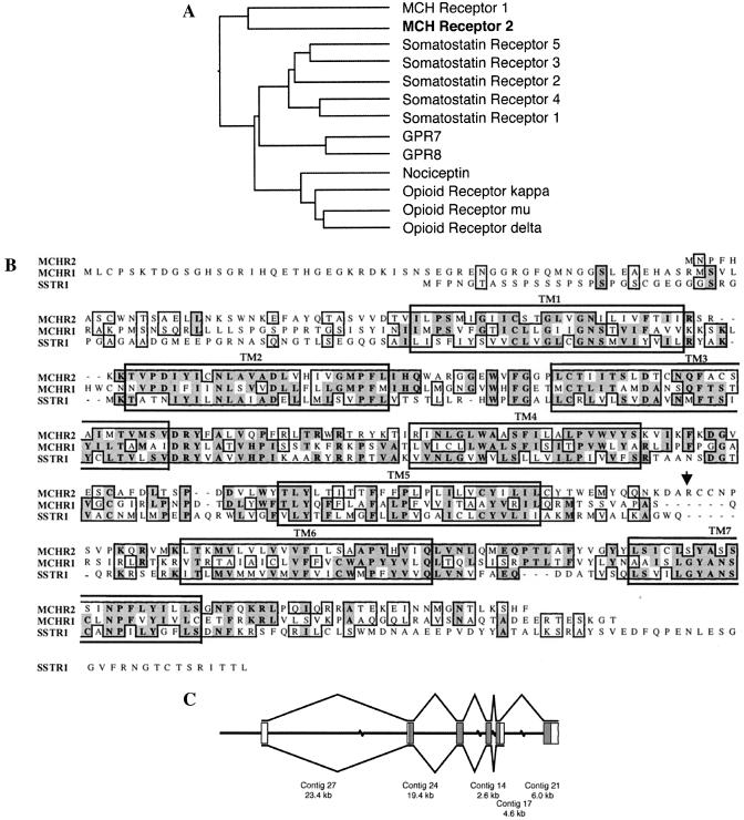
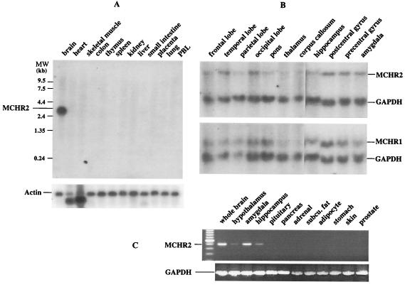
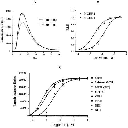
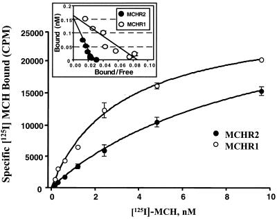
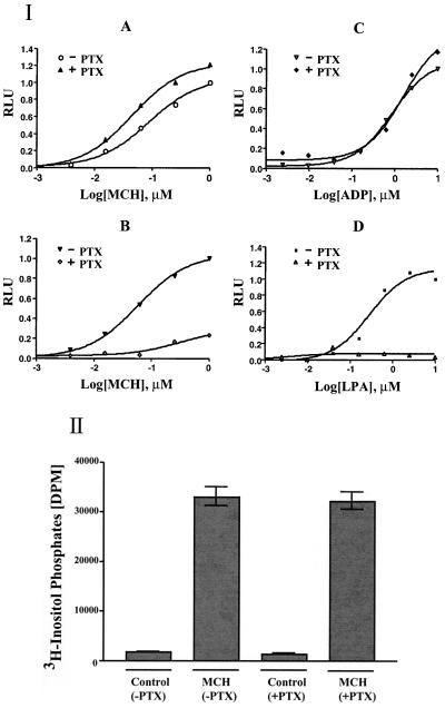
Melanin-concentrating hormone (MCH), a neuropeptide expressed in central and peripheral nervous systems, plays an important role in the control of feeding behaviors and energy metabolism. An orphan G protein-coupled receptor (SLC-1/GPR24) has recently been identified as a receptor for MCH (MCHR1). We report here the identification and characterization of a G protein-coupled receptor as the MCH receptor subtype 2 (MCHR2). MCHR2 has higher protein sequence homology to MCHR1 than any other G protein-coupled receptor. The expression of MCHR2 has been detected in many regions of the brain. In contrast to MCHR1, which is intronless in the coding region and is located at the chromosomal locus 22q13.3, the MCHR2 gene has multiple exons and is mapped to locus 6q21. MCHR2 is specifically activated by nanomolar concentrations of MCH, binds to MCH with high affinity, and signals through Gq protein. This discovery is important for a full understanding of MCH biology and the development of potential therapeutics for diseases involving MCH, including obesity.


Figures

Figure 1
Figure 1
Analysis of the sequences, genomic structure, and splicing of the MCHR2 gene. (A) Sequence similarity between MCHR2 and related GPCRs. The protein sequence of MCHR2 was aligned with the GPCRs most similar to it, including all of the members of the somatostatin and opioid receptor families and two orphan GPCRs, GPR7 and GPR8. The GCG programs distances and growtree were used to generate a similarity tree based on the alignment. The lengths of the horizontal line segments connecting two GPCRs are inversely proportional to the similarities between their protein sequences. (B) Alignment of the protein sequences of MCHR1, MCHR2, and SSTR1. Identical and similar amino acid residues are boxed, with identical ones in shadowed boxes. The large boxes indicate predicted transmembrane domains. The arrow indicates the end of the putative truncated form of the MCHR2 protein as a result of alternative splicing. (C) MCHR2 exon structure. The MCHR2 mRNA sequence was used to order and orient five of the unordered contigs in the GenBank BAC sequence AC027643. The central line represents the BAC sequence, with discontinuities representing gaps of unknown length between contigs. Boxes represent regions of the genomic sequence containing MCRH2 exons; filled boxes are translated, and open boxes contain 5′ and 3′ untranslated region sequences. Lines above and below the exons indicate the two splicing patterns observed. The position of the 3′ end of the 3′ untranslated region of the longer transcript is unknown. The exon lengths are drawn out of scale with the contig lengths.
Figure 2
Figure 2
MCHR2 expression in the brain. (A) Northern hybridization of the human multiple tissue mRNA blot. MCHR2 expression was detected only in the brain. (B) Northern hybridization of human brain blots. MCHR2, like MCHR1, was present in many brain regions.
Figure 3
Figure 3
MCHR2 is a receptor for MCH. (A) MCH stimulates a transient increase in intracellular Ca2+ similar to that resulting from MCH activation of MCHR1. CHO cells were transfected with MCHR2 or MCHR1 expression plasmids. The intracellular Ca2+ levels were measured with an aequorin luminescent assay (Materials and Methods). (B) MCH activated MCHR2 with an EC50 comparable to that observed with MCHR1 in transiently transfected HEK293 cells. (C) Specific activation of MCHR2 by MCH. MCH or MCH-related peptides were tested for activation of MCHR2 receptor by the aequorin assay. Only MCH, salmon MCH, and the MCH analog [Phe13, Tyr19]-MCH [MCH (P/T)] induced a Ca2+ increase in MCHR2-transfected HEK293 cells. SST14, somatostatin-14; CS14, cortistatin-14; αMSH, α-melanotropin; NEI, neuropeptide-EI (MCH-precursor-derived peptide).
Figure 4
Figure 4
Binding of 125I-labeled MCH to MCHR2. Radioligand binding was performed as described in Materials and Methods. Binding of MCHR1 to 125I-labeled MCH was also performed for comparison. Smooth lines are a nonlinear regression fit. MCHR2 bound to MCH with an affinity (Kd = 9.6 ± 0.5 nM) comparable to that of MCHR1 (Kd = 3.1 ± 0.4 nM). (Inset) Scatchard plots. The Bmax was about 0.16 nM for both MCHR2 and MCHR1, corresponding to 1.6 nmol/g membrane protein. Data are expressed as mean ± SD (n = 3).
Figure 5
Figure 5
MCHR2 is primarily coupled to Gq. (I) HEK293 cells transfected with MCHR2 or MCHR1 were stimulated with MCH, ADP, or lysophosphatidic acid (LPA) at the indicated concentrations with or without pretreatment with PTX (100 ng/ml) for 16 h. The Ca2+ responses were measured with the aequorin assay. Relative luminescence (RLU) was the ratio of luminescence units from cells with different treatments to that from cells treated with the highest concentration of ligand in the absence of PTX. The cells were transfected with MCHR2 (A) or MCHR1 (B) and stimulated with MCH. (C and D) HEK293 cells transiently transfected with MCHR2 were also stimulated with ADP (C) or LPA (D) to activate their endogenous receptors in the cells. (II) MCH increased the production of inositol phosphates in the MCHR2-transfected cells, and this effect is insensitive to PTX. HEK293 cells transfected with MCHR2 were stimulated with 100 nM MCH with or without PTX. Controls did not receive MCH.

References

    1. Kawauchi H, Kawazoe I, Tsubokawa M, Kishida M, Baker B I. Nature (London) 1983;305:321–323. - PubMed
    1. Skofitsch G, Jacobowitz D M, Zamir N. Brain Res Bull. 1985;15:635–649. - PubMed
    1. Bittencourt J C, Presse F, Arias C, Peto C, Vaughan J, Nahon J L, Vale W, Sawchenko P E. J Comp Neurol. 1992;319:218–245. - PubMed
    1. Saito Y, Nothacker H-P, Civelli O. Trends Endocrinol Metabol. 2000;11:299–303. - PubMed
    1. Qu D, Ludwig D S, Gammeltoft S, Piper M, Pelleymounter M A, Cullen M J, Mathes W G, Przypek J, Kanarek R, Maratos-Flier E. Nature (London) 1996;380:243–247. - PubMed

MeSH terms

LinkOut - more resources