. 2001 Jul;183(14):4115-26.
doi: 10.1128/JB.183.14.4115-4126.2001.

Constitutive septal murein synthesis in Escherichia coli with impaired activity of the morphogenetic proteins RodA and penicillin-binding protein 2

Affiliations

Constitutive septal murein synthesis in Escherichia coli with impaired activity of the morphogenetic proteins RodA and penicillin-binding protein 2

M A de Pedro et al. J Bacteriol. 2001 Jul.

Abstract

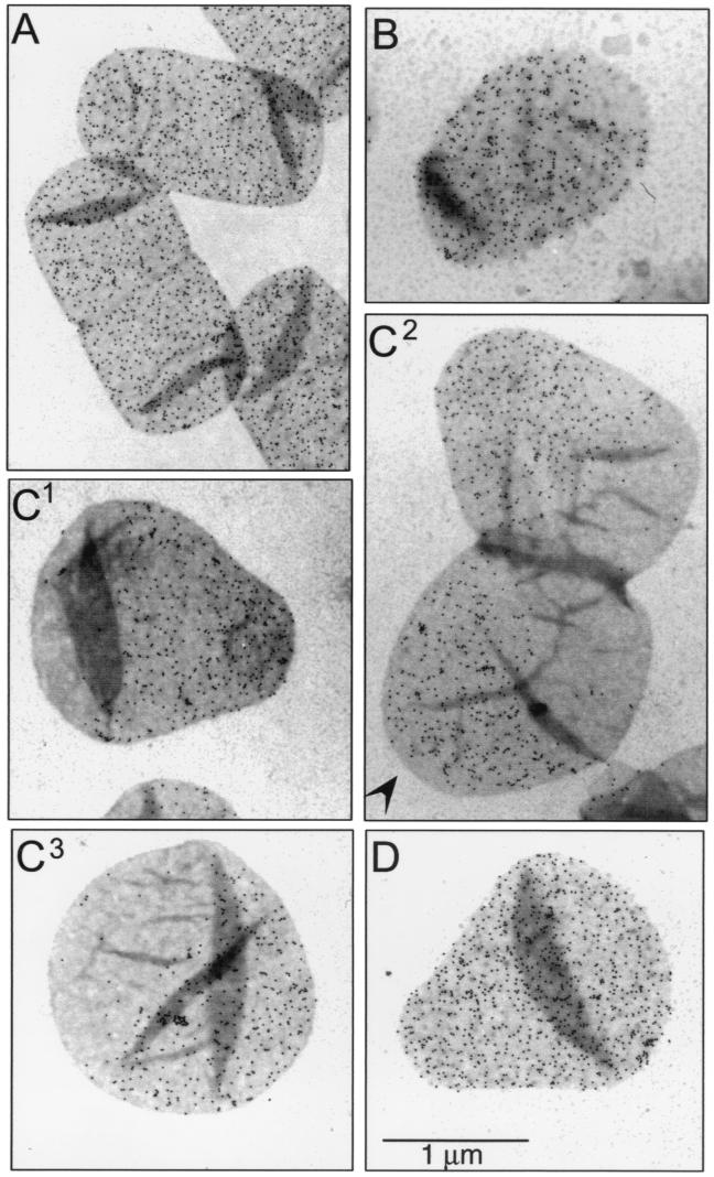
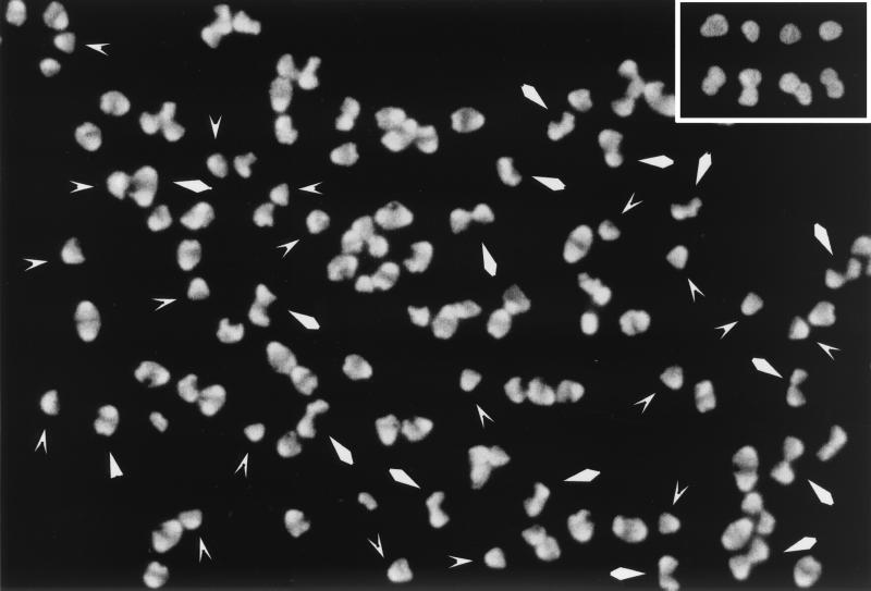
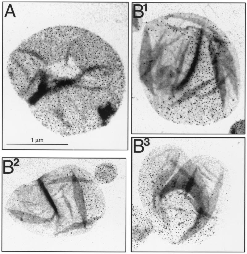
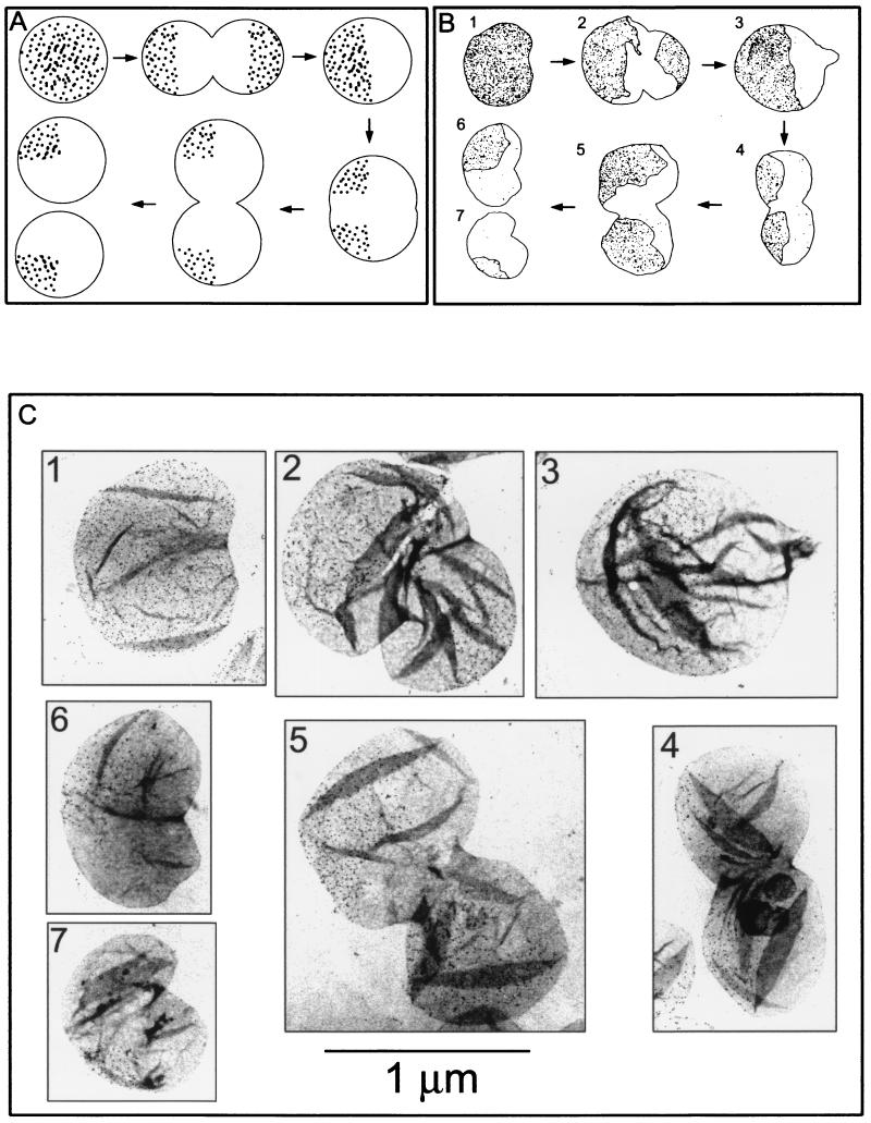
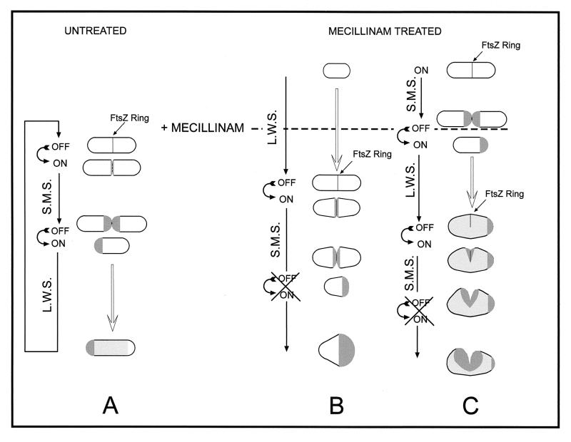
The pattern of peptidoglycan (murein) segregation in cells of Escherichia coli with impaired activity of the morphogenetic proteins penicillin-binding protein 2 and RodA has been investigated by the D-cysteine-biotin immunolabeling technique (M. A. de Pedro, J. C. Quintela, J.-V. Höltje, and H. Schwarz, J. Bacteriol. 179:2823-2834, 1997). Inactivation of these proteins either by amdinocillin treatment or by mutations in the corresponding genes, pbpA and rodA, respectively, leads to the generation of round, osmotically stable cells. In normal rod-shaped cells, new murein precursors are incorporated all over the lateral wall in a diffuse manner, being mixed up homogeneously with preexisting material, except during septation, when strictly localized murein synthesis occurs. In contrast, in rounded cells, incorporation of new precursors is apparently a zonal process, localized at positions at which division had previously taken place. Consequently, there is no mixing of new and old murein. Old murein is preserved for long periods of time in large, well-defined areas. We propose that the observed patterns are the result of a failure to switch off septal murein synthesis at the end of septation events. Furthermore, the segregation results confirm that round cells of rodA mutants do divide in alternate, perpendicular planes as previously proposed (K. J. Begg and W. D. Donachie, J. Bacteriol. 180:2564-2567, 1998).


Figures

FIG. 1
FIG. 1
Immunoelectron microscopy of d-Cys-labeled murein in amdinocillin-treated cells of MC6RP1. Cells from a culture grown for three generations in LB medium supplemented with d-Cys were harvested by centrifugation and transferred to medium without d-Cys, but containing 1 μg of amdinocillin per ml. As a control, an aliquot was transferred to medium with both amdinocillin and d-Cys at the concentrations shown above. At the indicated chase times, samples were removed and further processed for murein purification, biotinylation, immunolabeling, and electron microscopy as described in Materials and Methods. Gold-conjugated (6-nm-diameter grains) protein A was used for the detection of antibodies. (A) Chase time zero. (B) Cells chased for one doubling in cell mass. (C) Cells chased for two doublings in cell mass. The arrowhead indicates a gold-free pole. (D) Control cells incubated for two doublings in cell mass in d-Cys plus amdinocillin. All pictures are at the same magnification.
FIG. 2
FIG. 2
Immunofluorescence microscopy of d-Cys-labeled murein in amdinocillin-treated cells of MC6RP1. Aliquots of the same sacculi purified for the experiment shown in Fig. 1 were subjected to immunofluorescence microscopy for the detection of d-Cys-labeled areas with a fluorescein isothiocyanate-labeled goat anti-rabbit antibody. The picture shows sacculi chased for two doublings in cell mass, corresponding to Fig. 1C. V-shaped and rhomboidal arrowheads indicate sacculi similar to the ones in Fig. 1C1 and C2, respectively. The inset shows selected sacculi from the sample incubated for the same time in d-Cys plus amdinocillin (Fig. 1D).
FIG. 3
FIG. 3
Immunoelectron microscopy of d-Cys-labeled murein in cells of the pbpA(Ts) mutant SP4500. Cells from a culture grown for three generations in LB medium supplemented with d-Cys at 42°C were harvested by centrifugation. One sample was immediately subjected to murein purification, and the rest of the cell suspension was transferred to prewarmed medium without d-Cys and further incubated for one and a half generations. Sacculi were purified, biotinylated, and immunolabeled as indicated in Materials and Methods. Gold-conjugated (6-nm-diameter grains) protein A was used for the detection of antibodies. (A) Nonchased sacculi. (B) Sacculi chased for one and a half doublings in mass. All pictures are at the same magnification.
FIG. 4
FIG. 4
Immunoelectron microscopy of d-Cys-labeled murein in cells of the rodA(Ts) mutant SP5211. Cells from a culture grown for three generations in LB medium supplemented with d-Cys (150 μg/ml) at 30°C were harvested and transferred to d-Cys-free medium prewarmed at 30 and 42°C. Cultures were further incubated at the respective temperature, and after one and two doublings in cell mass, samples were removed and further processed for sacculus purification, biotinylation, immunolabeling, and electron microscopy as indicated in Materials and Methods. Gold-conjugated (6-nm-diameter grains) protein A was used for the detection of antibodies. (A) Sacculi from control, nonchased cells. (B) Sacculi chased for one doubling in cell mass at 42°C. (C) Sacculi chased for two doublings in cell mass at 42°C. (D) Sacculi chased for two doublings in cell mass at 30°C. All pictures are at the same magnification.
FIG. 5
FIG. 5
Murein segregation in the rodA° mutant KJB24 as observed by immunoelectron microscopy. Cells from a culture grown for three generations in LB medium supplemented with d-Cys were harvested and transferred to prewarmed d-Cys-free medium. Cultures were further incubated, and after one and two doublings in cell mass, samples were removed and further processed for sacculus purification, biotinylation, immunolabeling, and electron microscopy as indicated in Materials and Methods. Gold-conjugated (6-nm-diameter grains) protein A was used for the detection of antibodies. (A) Hypothetical segregation pattern. (B) Schematic representation of the actual segregation pattern as found in the images shown in panel C. The profile, position, and number of dots were drawn from the real pictures by using digitized images and PhotoShop software. (C) Immunolabeling of d-Cys in selected sacculi after no chase (panel 1) or after chase for one (panels 2 and 3) and two (panels 4 to 7) doublings in cell mass. The numbers in panels B and C correspond. All pictures are at the same magnification.
FIG. 6
FIG. 6
Diagrammatic representation of the generation of rounded cells by the action of amdinocillin (mecillinam). Dark gray areas show newly synthesized septal murein; Light gray areas show newly made lateral wall murein. L.W.S., lateral wall murein synthesis.

Comment in

References

    1. Addinall S G, Lutkenhaus J. FtsZ-spirals and -arcs determine the shape of the invaginating septa in some mutants of Escherichia coli. Mol Microbiol. 1996;22:231–237. - PubMed
    1. Ayala J A, Garrido T, de Pedro M A, Vicente M. Molecular biology of bacterial septation. In: Ghuysen J M, Hakenbeck R, editors. Bacterial cell wall. Amsterdam, The Netherlands: Elsevier; 1994. pp. 73–101.
    1. Begg K J, Donachie W D. Cell shape and division in Escherichia coli: experiments with shape and division mutants. J Bacteriol. 1985;163:615–622. - PMC - PubMed
    1. Begg K J, Donachie W D. Division planes alternate in spherical cells of Escherichia coli. J Bacteriol. 1998;180:2564–2567. - PMC - PubMed
    1. Begg K J, Spratt B G, Donachie W D. Interaction between membrane proteins PBP3 and RodA is required for normal cell shape and division in Escherichia coli. J Bacteriol. 1986;167:1004–1008. - PMC - PubMed

Publication types

MeSH terms