. 2001 Jul;183(14):4317-22.
doi: 10.1128/JB.183.14.4317-4322.2001.

Localization of GerAA and GerAC germination proteins in the Bacillus subtilis spore

Affiliations

Localization of GerAA and GerAC germination proteins in the Bacillus subtilis spore

K D Hudson et al. J Bacteriol. 2001 Jul.

Abstract

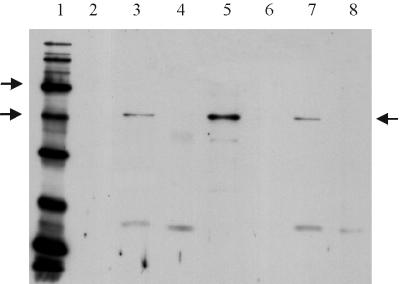
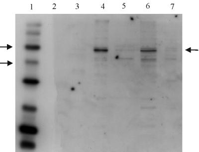
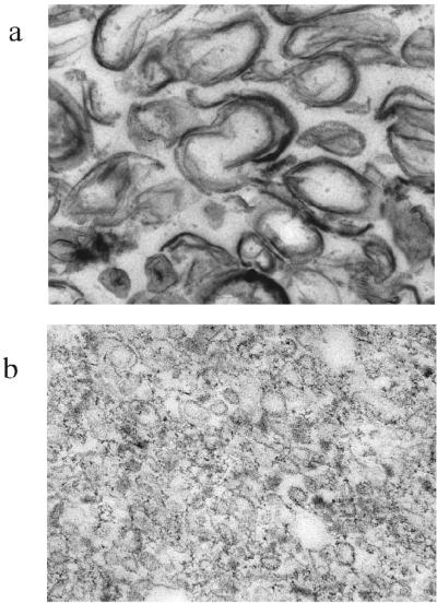
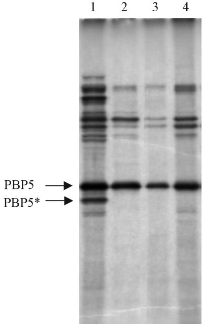
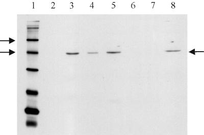
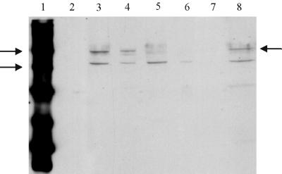
The GerAA, -AB, and -AC proteins of the Bacillus subtilis spore are required for the germination response to L-alanine as the sole germinant. They are likely to encode the components of the germination apparatus that respond directly to this germinant, mediating the spore's response; multiple homologues of the gerA genes are found in every spore former so far examined. The gerA operon is expressed in the forespore, and the level of expression of the operon appears to be low. The GerA proteins are predicted to be membrane associated. In an attempt to localize GerA proteins, spores of B. subtilis were broken and fractionated to give integument, membrane, and soluble fractions. Using antibodies that detect Ger proteins specifically, as confirmed by the analysis of strains lacking GerA and the related GerB proteins, the GerAA protein and the GerAC+GerBC protein homologues were localized to the membrane fraction of fragmented spores. The spore-specific penicillin-binding protein PBP5*, a marker for the outer forespore membrane, was absent from this fraction. Extraction of spores to remove coat layers did not release the GerAC or AA protein from the spores. Both experimental approaches suggest that GerAA and GerAC proteins are located in the inner spore membrane, which forms a boundary around the cellular compartment of the spore. The results provide support for a model of germination in which, in order to initiate germination, germinant has to permeate the coat and cortex of the spore and bind to a germination receptor located in the inner membrane.


Figures

FIG. 1
FIG. 1
Proteins in extracts from broken and fractionated spores were separated on an SDS–12% polyacrylamide gel and transferred to PVDF membrane for Western blotting by the Amersham ECL chemiluminescence detection system. Each sample track contains ca. 11 μg of protein. Detection of GerAC was achieved using anti-GerAC antibody, diluted at 1/500. The secondary antibody was used at 1/15,000 dilution. Lane 1 contained biotinylated protein standards (Sigma SDS-6B: sizes of 205, 116, 97, 58, 40, 29, 20, 14.3, and 6.5 kDa). Arrows by the marker lane indicate the 58 and 40-kDa markers, and the arrow on the right of the gel indicates the GerAC band. Lane 2 contained prestained protein markers, lane 3 contained total extract from broken spores of strain 1604, and lanes 4 to 6 contained integument, membrane, and soluble fractions, respectively, of strain 1604. Lanes 7 and 8 contained total spore extracts of strains AM418 (gerAΔ) and AM1422 (gerAΔ gerB null), respectively.
FIG. 2
FIG. 2
Western blot of spore extracts probed with anti-GerAA anti-peptide antibody. Conditions used were as for Fig. 1, except that the secondary antibody was used at a 1 in 10,000 dilution. Lanes 1 and 2 contained biotinylated and prestained protein markers, respectively. Arrows by the marker lane indicate the 58 and 40-kDa markers, and the arrow on the right of the gel indicates the GerAA band. Lanes 3 to 5 contained integument, membrane, and soluble fractions, respectively, of dormant spores of strain WB600. Lane 6 contains total extract from WB600 spores, and lane 7 contained an extract of spores of strain AM1422 (gerAΔ gerB null).
FIG. 3
FIG. 3
Electron micrographs of material in integument (a) and membrane (b) fractions. Magnification: ×21,000 and ×78,000, respectively.
FIG. 4
FIG. 4
Penicillin-binding protein profiles. Penicillin-binding proteins were labeled with [3H]penicillin and separated on an SDS–10% polyacrylamide gel. Bands corresponding to PBP5 and PBP5∗ are arrowed. Lane 1, total spore membranes from AM047; lane 2, the membrane fraction from dormant spores of strain 1604; lane 3, the membrane fraction from germinated spores of 1604, as used for Western blotting; lane 4, membrane from coat-stripped spores of 1604. The autoradiograph has been overdeveloped to expose faint bands to establish that there is no evidence of PBP5∗ in the membrane fractions in lanes 2 and 3.
FIG. 5
FIG. 5
Western blot of GerAC and GerBC proteins from coat-stripped spores. Each sample track contains ca. 11 μg of protein. Conditions for electrophoresis and Western blotting were as for Fig. 1. Arrows by the marker lane indicate the 58- and 40-kDa markers, and the arrow on the right of the gel indicates the GerAC band. Lane 1, biotinylated markers; lane 2, prestained protein markers; lane 3, total spore extract of strain WB600; lane 4, total spore extract of strain AM047 (gerE36). Lanes 5 and 6 contained total spore extracts of the gerAΔ and gerAΔ gerBΔ strains respectively. Lane 7 contains concentrated extracted coat and outer membrane material from spores of strain AM047, and lane 8 contains protein from the broken, decoated, AM047 spores.
FIG. 6
FIG. 6
Detection of GerAA protein in Western blots of coat-stripped spores. Lanes 3 to 9 contain 11 μg of protein. Conditions were as described for Fig. 2. Lanes 1 and 2 are markers, as in the other Western blots. Arrows by the marker lane indicate the 58- and 40-kDa biotinylated protein standards in Sigma SDS-6B, and the arrow on the right indicates the position of the GerAA band. Lane 3 and 4 contained total spore extracts of WB600 and AM047, respectively. Lanes 5 and 6 contained extracts from the gerA and gerAB null mutant spores, respectively. Lane 7 contains concentrated extracted coat and outer membrane material from spores of strain AM047, and lane 8 contains protein from the broken, decoated, AM047 spores.

References

    1. Behravan J, Chirakkal H, Masson A, Moir A. Mutations in the gerP locus of Bacillus subtilis and Bacillus cereus affect the access of germinants to their target in the spore. J Bacteriol. 2000;182:1987–1994. - PMC - PubMed
    1. Buchanan C E, Neyman S L. Correlation of penicillin binding protein composition with different functions of two membranes in Bacillus subtilis forespores. J Bacteriol. 1986;165:498–503. - PMC - PubMed
    1. Chambers S P, Prior S E, Barstow D A, Minton N. The pMTL nic-cloning vectors. I. Improved pUC polylinker regions to facilitate the use of sonicated DNA for nucleotide sequencing. Gene. 1988;68:139–149. - PubMed
    1. Clements M O, Moir A. Role of the gerI operon of Bacillus cereus 569 in the response of spores to germinants. J Bacteriol. 1998;180:6729–6735. - PMC - PubMed
    1. Corfe B M, Moir A, Popham D, Setlow P. Analysis of the expression and regulation of the gerB spore germination operon of Bacillus subtilis 168. Microbiology. 1994;140:3079–3083. - PubMed

Publication types