. 2001 Jul;65(1):318-32.
doi: 10.1095/biolreprod65.1.318.

Spermatogenesis in Bclw-deficient mice

Affiliations

Spermatogenesis in Bclw-deficient mice

L D Russell et al. Biol Reprod. 2001 Jul.

Abstract

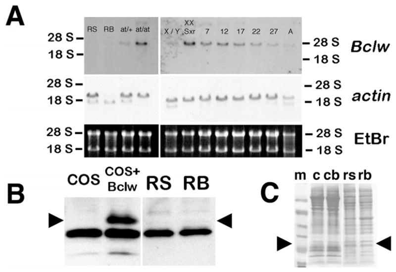
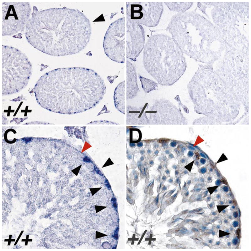
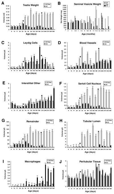
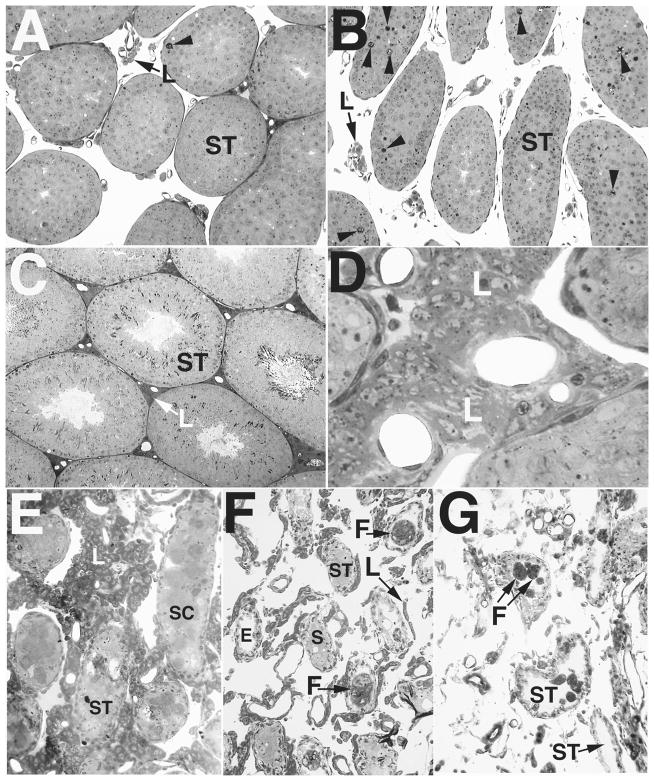
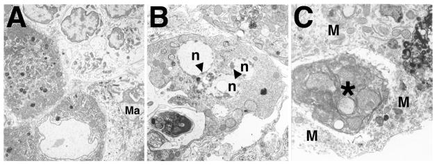
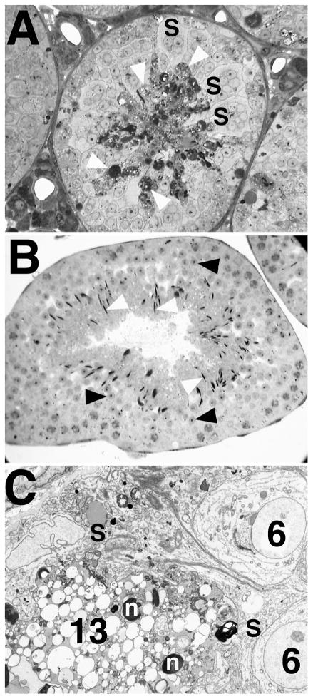
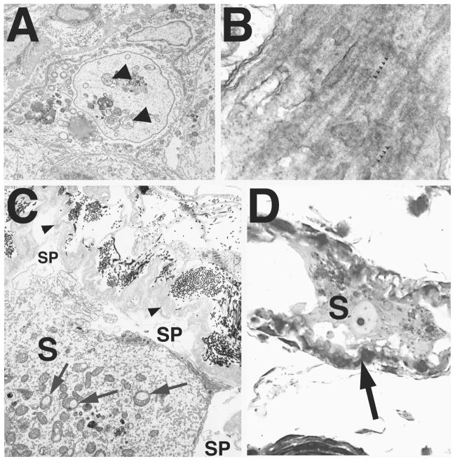
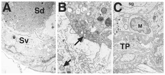
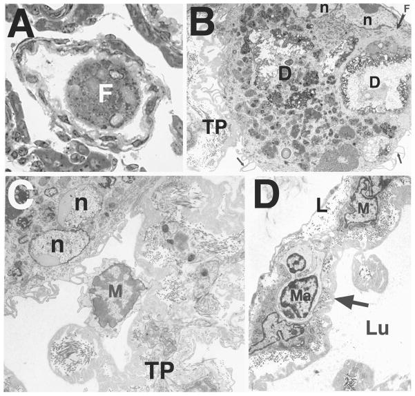
Bclw is a death-protecting member of the Bcl2 family of apoptosis-regulating proteins. Mice that are mutant for Bclw display progressive and nearly complete testicular degeneration. We performed a morphometric evaluation of testicular histopathology in Bclw-deficient male mice between 9 days postnatal (p9) through 1 yr of age. Germ cell loss began by p22, with only few germ cells remaining beyond 7 mo of age. A complete block to elongated spermatid development at step 13 occurred during the first wave of spermatogenesis, whereas other types of germ cells were lost sporadically. Depletion of Sertoli cells commenced between p20 and p23 and continued until 1 yr of age, when few, if any, Sertoli cells remained. Mitochondria appeared to be swollen and the cytoplasm dense by electron microscopy, but degenerating Bclw-deficient Sertoli cells failed to display classical features of apoptosis, such as chromatin condensation and nuclear fragmentation. Macrophages entered seminiferous tubules and formed foreign-body giant cells that engulfed and phagocytosed the degenerated Sertoli cells. Leydig cell hyperplasia was evident between 3 and 5 mo of age. However, beginning at 7 mo of age, Leydig cells underwent apoptosis, with dead cells being phagocytosed by macrophages. The aforementioned cell losses culminated in a testis-containing vasculature, intertubular phagocytic cells, and peritubular cell "ghosts." An RNA in situ hybridization study indicates that Bclw is expressed in Sertoli cells in the adult mouse testis. Consequently, the diploid germ cell death may be an indirect effect of defective Sertoli cell function. Western analysis was used to confirm that Bclw is not expressed in spermatids; thus, loss of this cell type most likely results from defective Sertoli cell function. Because Bclw does not appear to be expressed in Leydig cells, loss of Leydig cells in Bclw-deficient mice may result from depletion of Sertoli cells. Bclw-deficient mice serve as a unique model to study homeostasis of cell populations in the testis.


Figures

FIG. 1
FIG. 1
Expression of Bclw mRNA and protein in male mouse germ cells. A) RNA was extracted from preparations of enriched germ cells, from testes of heterozygous or homozygous mutant atrichosis (at) mice, from testes of normal (X/Y) or sex-reversed mice (XX Sxr), or from testes of mice at Postnatal Day 7, 12, 17, 22, and 27 during prepubertal development and from adults. The percentage of contaminants in the enriched germ cell preparations is as described in Materials and Methods. Testes from at/at and XX Sxr animals contain extremely few or no germ cells, respectively; thus, any signal is derived from somatic cells of the testis. The RNAs were subjected to Northern blot analysis using a full-length Bclw cDNA probe. To determine whether equal amounts of total RNA had been loaded, the gel was stained with ethidium bromide (EtBr), photographed under ultraviolet (UV) light before transfer, and the blots hybridized with an actin cDNA (lower and middle panels, respectively). A, Testes from adult mouse; RB, residual bodies; RS, round spermatids; at/+, testis from heterozygote atrichosis mouse; at/at, testis from homozygote atrichosis mouse; X/Y, testis from wild-type mouse; XX Sxr, testis from sex-reversed mouse; 7, 12, 17, 22, and 27, testes from Postnatal Day 7, 12, 17, 22, and 27 mice. B) Western blot analysis of total protein in preparations of enriched germ cells. Cell preparations enriched for round spermatids (RS) or residual bodies (RB) from adult animals were solubilized, and total protein was analyzed by Western blot analysis using affinity-purified rabbit antisera against a peptide sequence from mouse Bclw [5]. The positive and negative controls include extracts of COS cells transfected with either a mammalian expression vector with a mouse Bclw cDNA insert (COS + Bclw) or the same vector without a cDNA insert (COS). Arrowheads indicate the position of the Bclw-specific band. Note the absence of Bclw protein in both RS and RB. C) To determine whether equal amounts of protein had been loaded, the blot shown in B was stained with Ponceau S before reacting with antisera. Arrowheads denote the approximate size of Bclw (22 kDa). c, COS cells; cb, COS cells transfected with vector expressing Bclw; m, molecular weight markers; rb, enriched for residual bodies; rs, enriched for round spermatids.
FIG. 2
FIG. 2
In situ hybridization analysis of expression of Bclw mRNA in mouse testis. A) Section of testis from adult wild-type mouse hybridized with an antisense ribo-probe derived from the 3′-UTR of Bclw cDNA. A significant positive signal (a purple precipitate) is observed only over nuclei of Sertoli cells. Significant signal was observed in all stages of tubules, except those between stages VII and VIII of the seminiferous cycle (arrowhead). ×200. B) Section of testis from adult Bclw homozygous mutant prepared and analyzed simultaneously under identical conditions as those in A. Only background purple precipitate is seen. ×200. C) Higher magnification of a single seminiferous tubule from a wild-type mouse, hybridized with anti-sense Bclw 3′-UTR riboprobe. Sertoli cell nuclei (black arrowheads) stain relatively intensely purple. In contrast, a spermatogonium (red arrowhead) does not display any significant staining over background. ×640. D) Serial section from that shown in C reacted with antibody for GATA-1, a marker of Sertoli cell nuclei. The brown staining denotes presence of the antibody. The blue counterstain is hematoxylin. Black and red arrowheads denote the same cells as identified in C. ×640.
FIG. 3
FIG. 3
Quantitative and morphometric analysis of testicular composition in Bclw mutant and control mice. Except for B, in each graph the wild-type (WT) and heterozygous mutant (Het) animals are pooled, because no significant difference was found in testicular morphology between these two genotypes. No significant difference was found in weights of seminal vesicles between WT and Het animals, but the values in B are presented separately, with each value representing an individual animal. For the remaining graphs, an asterisk indicates that only two animals were in these groups, hence the omission of a bar denoting SEM.
FIG. 4
FIG. 4
Light micrographs showing development of the mutant testicular phenotype in Bclw-deficient mice, emphasizing the general appearance of the testis and the Leydig cells. A and B) Testes at 20 days of age showing the similar appearance of control (A) and Bclw-mutant animals (B) with respect to Leydig cells (L). As previously described [5], approximately sevenfold more degenerating cells (arrowheads) are seen in the seminiferous tubules (ST) from mutants than in the control. ×70 and ×60, respectively. C and D) Testes from 3-mo-old animals showing development of the Leydig cells (L) in control (C) and Bclw-mutant animals (D). Intertubular clusters of Leydig cells are extensive in Bclw mutants. ×80 and ×350, respectively. E) Testes from 5.5-mo-old mutants showing the prominent presence of Leydig cells (L). Large masses in seminiferous tubules (ST) were composed of sloughed dead and or dying Sertoli cells (SC). ×155. F) Testes from 8-mo-old mutants showing small “strings” of Leydig cells (L) among extremely small seminiferous tubules (ST), with the latter containing some Sertoli cells (S) or foreign-body giant cells (F). A seminiferous tubules devoid of cellular contents (peritubular “ghosts”) is marked (E). ×75. G) Testes from 12-mo-old mutants with absence of Leydig cells. The scattered cells between peritubular “ghosts” are connective tissue elements and phagocytic elements. A few foreign-body giant cells (F) are seen within seminiferous tubules (ST). ×100.
FIG. 5
FIG. 5
Ultrastructural appearance of Leydig cells in Bclw-mutants. A) At 34 days of age, Leydig cells (darker cells in the lower left) appeared normal. Often, a mononuclear infiltrate was seen in the interstitium (lighter cells in the upper right). A macrophage is also indicated (Ma). ×4500. B) Beginning around 8 mo of age, Leydig cells in mutants were observed undergoing apoptosis, as evidenced by the fragmented nuclei (n) and accumulation of associated peripheral heterochromatin (arrowheads). The Leydig cell could be recognized as such by the abundant smooth endoplasmic reticulum and mitochondria possessing tubular cristae and by comparison with relatively normal-appearing, adjacent Leydig cells (top right). ×7200. C) Phagocytosis of degenerating Leydig cell (asterisk) by an interstitial macrophage (M). The phagocytosed cell appears dense and contains mitochondria characteristic of those found in Leydig cells. ×14 000.
FIG. 6
FIG. 6
Degeneration of elongate spermatids in Bclw-deficient mice. A) Light micrograph showing degeneration of step 13 spermatids (white arrowheads), appearing as very dense material, in seminiferous tubules from a Bclw homozygous mutant. Symplasts of round spermatids (S) were also commonly seen in mutants. ×275. B) Light micrograph showing a comparable stage I tubule in a control animal. Normally developing round (black arrowheads) and elongate (white arrowheads) spermatids are marked. ×170. C) Electron micrograph of a stage VI seminiferous tubule from a Bclw-deficient animal. Step 6 (6) spermatids are associated with a dense, degenerating symplast of step 13 (13) spermatids (lower left) that is contained in a single, dense cytoplasmic mass in contact with Sertoli cells (S). Step 13 spermatid nuclei (n) are indicated. ×3200.
FIG. 7
FIG. 7
Morphological features of Sertoli cells in Bclw-deficient animals. A) Nuclear vacuolation (arrowheads) within the nucleus of a Sertoli cell located centrally within the micrograph. Many mitochondria appear swollen. ×3300. B) En face section of Sertoli-Sertoli junctions in p90 Bclw-deficient animal showing both actin filament bundles (gray parallel amorphous material running from lower left to upper right) and translucent linearities (small black arrowheads) in the areas of occluding junction representing fusions of membranes. ×85 000. C) Loss of attachment of Sertoli cells from the seminiferous tubule and from each other in a Bclw-mutant more than 6 mo of age. Spaces where the Sertoli cell (S) has not maintained contact with the highly infolded and duplicated basal lamina (arrowheads) are indicated (SP). No other Sertoli cell was seen at the lateral borders of this Sertoli cell (not shown). Many mitochondria in the Sertoli cytoplasm appear swollen (arrows). ×10 000. D) Light micrograph of a single Sertoli cell (S) in a greatly shrunken tubule from a Bclw mutant more than 6 mo of age containing only peritubular cells of the tunica propria (arrow). No other Sertoli cell was seen at its lateral borders. Note the vacuolation within the Sertoli cell cytoplasm. No Leydig cells are shown. ×1200.
FIG. 8
FIG. 8
Electron micrographs illustrating degenerating Sertoli cell masses located centrally within seminiferous tubules in 9-mo-old Bclw mutants. A) A large, dense Sertoli cell mass (Sd) is located in the center of a germ cell-depleted tubule adjacent to a viable Sertoli cell (Sv). ×4000. B) The interface of a highly densified, dying or dead Sertoli cells (upper right) with normal-appearing, viable Sertoli cells (lower left) shows the mitochondria of both to be characteristic, mouse-type Sertoli cell mitochondria (arrows). ×18 000. C) Inactivated macrophage (M) within the seminiferous tubule among Sertoli cells and a spermatogonium (sg). The tunica propria (TP) of the tubule is at the bottom of the figure. ×5500.
FIG. 9
FIG. 9
Formation of foreign-body giant cells in seminiferous epithelium of Bclw-mutants. A) Light micrograph showing a foreign-body giant cell (F) in the seminiferous tubules of a Bclw-deficient mouse. ×1100. B) Electron micrograph of a foreign-body giant cell. These extremely large, phagocytic foreign-body giant cells (arrow, F) contain phagocytosed material and were seen primarily in tubules depleted of cells, with only the peritubular cells of the tunica propria (TP) at the left remaining. Ultrastructurally, foreign-body giant cells display multiple nuclei (n), microvillous processes (small arrows at bottom), and an abundance of semidigested cellular remnants (D) contained within residual bodies. ×5000. C) Seminiferous tubule with only a tunica propria (TP) lining and intratubular macrophages (M). Both an inactivated macrophage (M) and a portion of a foreign-body giant cell showing multiple nuclei (n) are seen. ×5000. D) Electron micrograph of the peritubular cell tunica propria forming the wall of a seminiferous tubule lacking cellular contents. Identified are the tubular lumen (Lu), the myoid cell (M), a thin and lymphatic endothelial cell (L), and the infolded and duplicated basal lamina (arrow) lying internal to the myoid cell. Also identified is a macrophage (Ma) within the tunica propria. ×6000.

References

    1. Capel B. The battle of the sexes. Mech Dev. 2000;92:89–103. - PubMed
    1. Vergouwen RPFA, Jacobs SGPM, Huiskamp R, Davids JAG, de Rooij DG. Proliferative activity of gonocytes, Sertoli cells and interstitial cells during testicular development in mice. J Reprod Fertil. 1991;93:233–243. - PubMed
    1. Steinberger A, Steinberger E. Replication pattern of Sertoli cells in maturing rat testis in vivo and in organ culture. Biol Reprod. 1971;4:84–87. - PubMed
    1. Russell LD, Bartke A, Goh JC. Postnatal development of the Sertoli cell barrier, tubular lumen, and cytoskeleton of Sertoli and myoid cells in the rat, and their relationship to tubular fluid secretion and flow. Am J Anat. 1989;184:179–189. - PubMed
    1. Ross A, Waymire KG, Moss JE, Parlow AF, Skinner MK, Russell LD, MacGregor GR. Testicular degeneration in Bclw-deficient mice. Nat Genet. 1998;18:251–255. - PubMed

Publication types

MeSH terms