

Review
doi: 10.1136/bmj.322.7301.1528.
Studies of apoptosis in breast cancer
Affiliations
- PMID: 11420276
- PMCID: PMC1120573
- DOI: 10.1136/bmj.322.7301.1528
Item in Clipboard
Review
Studies of apoptosis in breast cancer
BMJ.
.
No abstract available
Figures
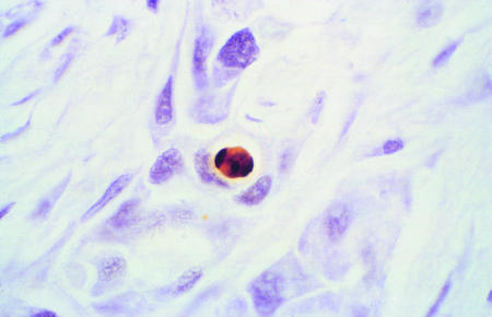
Histological sections of biopsy samples of breast tumours under light microscopy stained by TUNEL technique. Apoptotic cell with classic features of DNA condensation giving crescent shaped dense area and retraction of cytoplasm giving halo effect (top). Tumour necrosis showing accumulated nuclear debris and apoptotic bodies adjacent to invasive breast cancer (bottom). ×400

Histological sections of biopsy samples of breast tumours under light microscopy stained by TUNEL technique. Apoptotic cell with classic features of DNA condensation giving crescent shaped dense area and retraction of cytoplasm giving halo effect (top). Tumour necrosis showing accumulated nuclear debris and apoptotic bodies adjacent to invasive breast cancer (bottom). ×400
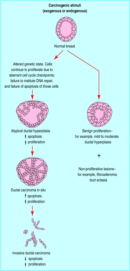
Phenotypic changes from normal breast, atypical ductal hyperplasia (intraepithelial neoplasia) through to intraductal carcinoma in situ and invasive ductal or lobular carcinoma
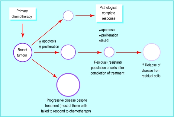
Schematic representation of tumours responding or resistant to chemotherapy and associated changes in growth control
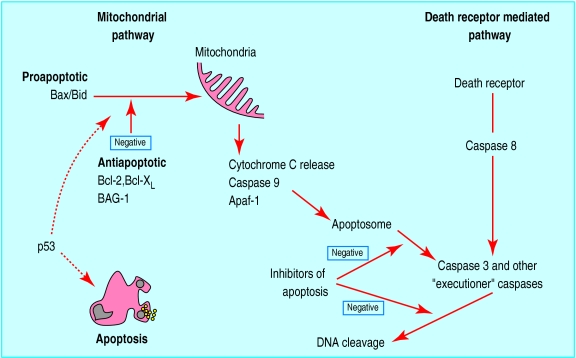
Relation between some major controlling factors involved in apoptosis. Two major pathways of caspase activation have been described in mammalian cells, resulting in apoptosis. In the mitochondrial pathway Bax and Bid are cell death factors, which increase mitochondrial permeability and release of cytochrome C. Cell survival factors Bcl-2, Bcl-XL, and BAG-1 inhibit actions of Bax and Bid on mitochondria. Apaf-1, a downstream mediator of apoptosis, along with cytochrome C, associates with caspase 9 in cytoplasm and leads to its activation. The resultant apoptosome initiates a cascade of effector caspases, which include caspases 3, 6, and 7. Active caspase 3 activates DNA fragmentation factor and promotes internucleosomal cleavage of DNA. The death receptor pathway is triggered by members of the death receptor superfamily such as CD95 and tumour necrosis factor receptor. Formation of a death inducing signalling complex induces caspase 8 activation and thereby the downstream caspase cascade. Further control of the apoptotic process is provided by inhibitors of apoptosis in the cytoplasm that abrogate caspase activity.Interaction between the two pathways exist. P53 is known to induce apoptosis, but exact mechanisms are not clear. The mitochondrial pathway seems to dominate in breast cancer
References
-
- Reed JC. Dysregulation of apoptosis in cancer. J Clin Oncol. 1999;17:2941–2953. - PubMed
-
- Tamm I, Schriever F, Dorken B. Apoptosis: implications of basic research for clinical oncology. Lancet Oncol. 2001;2:33–42. - PubMed
-
- Hickman JA. Apoptosis induced by anticancer drugs. Cancer Metastasis Rev. 1992;11:121–139. - PubMed
-
- Verheij M, Bartelink H. Radiation-induced apoptosis. Cell Tissue Res. 2000;301:133–142. - PubMed
Publication types
MeSH terms
Substances
LinkOut - more resources
Full Text Sources
Other Literature Sources
Medical