

Vascular differences detected by MRI for metastatic versus nonmetastatic breast and prostate cancer xenografts
- PMID: 11420750
- PMCID: PMC1505415
- DOI: 10.1038/sj.neo.7900129
Vascular differences detected by MRI for metastatic versus nonmetastatic breast and prostate cancer xenografts
Abstract
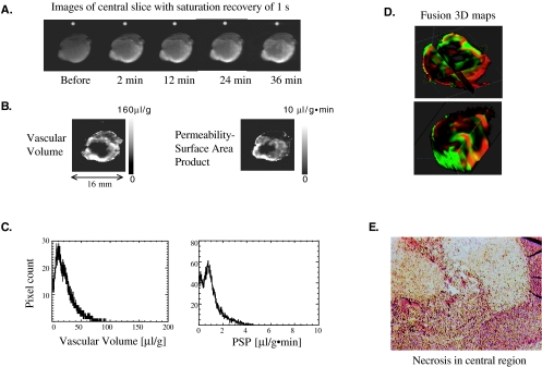
Several studies have linked vascular density, identified in histologic sections, to "metastatic risk." Functional information of the vasculature, not readily available from histologic sections, can be obtained with contrast-enhanced MRI to exploit for therapy or metastasis prevention. Our aims were to determine if human breast and prostate cancer xenografts preselected for differences in invasive and metastatic characteristics established correspondingly different vascular volume and permeability, quantified here with noninvasive MRI of the intravascular contrast agent albumin-GdDTPA. Tumor vascular volume and permeability of human breast and prostate cancer xenografts were characterized using MRI. Parallel studies confirmed the invasive behavior of these cell lines. Vascular endothelial growth factor (VEGF) expression in the cell lines was measured using ELISA and Western blots. Metastasis to the lungs was evaluated with spontaneous as well as experimental assay. Metastatic tumors formed vasculature with significantly higher permeability or vascular volume (P<.05, two-sided unpaired t test). The permeability profile matched VEGF expression. Within tumors, regions of high vascular volume usually exhibited low permeability whereas regions of low vascular volume exhibited high permeability. We observed that although invasion was necessary, without adequate vascularization it was not sufficient for metastasis to occur.
Figures

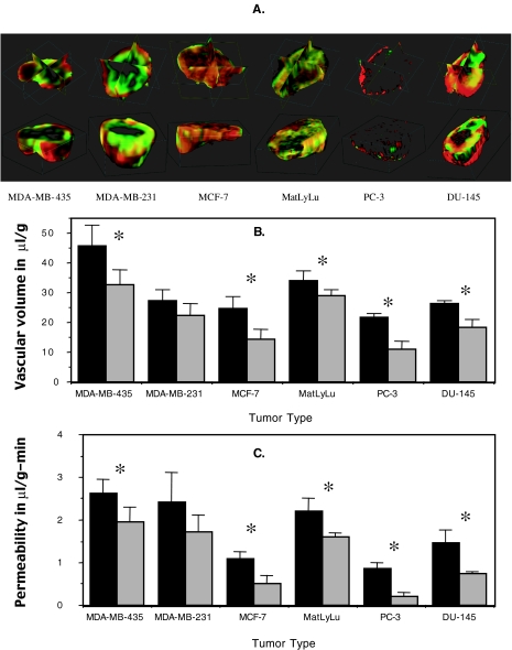
 ) are shown in (B). The mean value of the highest 25% of permeability values (■), and the permeability spatially corresponding to the highest 25% values of vascular volume (
 ) are shown in (C). A significant difference (P<.05) was observed for all the tumor models. The only exception to this was for the MDA-MB-231 tumor group.
References
-
- Weidner N, Semple JP, Welch WR, Folkman J. Tumor angiogenesis and metastasis-correlation in invasive breast carcinoma. N Engl J Med. 1991;324:1–8. - PubMed
-
- Thompson WD, Li WW, Maragoudakis M. The clinical manipulation of angiogenesis: pathology, side-effects, surprises, and opportunities with novel human therapies. J Pathol. 1999;187:503–510. - PubMed
-
- Horak ER, Leek R, Klenk N, LeJeune S, Smith K, Stuart N, Greenall M, Stepniewska K, Harris AL. Angiogenesis, assessed by platelet/endothelial cell adhesion molecule antibodies, as indicator of node metastases and survival in breast cancer. Lancet. 1992;340:1120–1124. - PubMed
-
- Wakui S, Furusato M, Itoh T, Sasaki H, Akiyama A, Kinoshita I, Asano K, Tokuda T, Aizawa S, Ushigome S. Tumour angiogenesis in prostatic carcinoma with and without bone marrow metastasis: a morphometric study. J Pathol. 1992;168:257–262. - PubMed
Publication types
MeSH terms
Substances
Grants and funding
LinkOut - more resources
Full Text Sources
Other Literature Sources
Medical