. 2001 Mar-Apr;3(2):154-64.
doi: 10.1038/sj.neo.7900128.

Suppression of angiogenesis and therapy of human colon cancer liver metastasis by systemic administration of interferon-alpha

Affiliations

Suppression of angiogenesis and therapy of human colon cancer liver metastasis by systemic administration of interferon-alpha

S Ozawa et al. Neoplasia. 2001 Mar-Apr.

Abstract

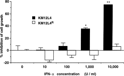
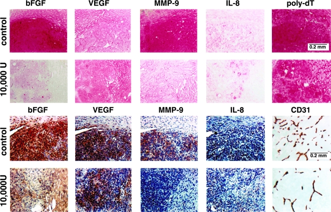
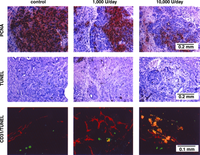
The purpose of this study was to determine whether systemic administration of interferon-alpha (IFN-alpha) can inhibit liver metastasis produced in nude mice by human colon cancer cells. KM12L4 (IFN-alpha-sensitive) or KM12L4 IFN(R) (IFN-alpha-resistant) cells were injected into the spleen of nude mice. Seven days later, the mice were treated with subcutaneous (s.c.) injections of IFN-alpha (70,000 units/week) at different dosing schedules (1, 2, or 7 times/week). Significant inhibition of tumor growth, vascularization and expression of basic fibroblast growth factor (bFGF) or matrix metalloproteinase-9 (MMP-9) mRNA and protein occurred in mice given daily injections of IFN-alpha. Kinetic analysis of therapy showed that daily s.c. administrations of 10,000 units of IFN-alpha induced apoptosis in liver metastasis-associated endothelial cells, followed by inhibition of tumor cell division and apoptosis of tumor cells. These data suggest that the antiangiogenic activity of IFN-alpha-2a depends on frequent administration of the optimal biologic dose.


Figures

Figure 1
Figure 1
Antiproliferative effects of IFN-α-2a against KM12L4 and KM12L4 IFNR cells. Tumor cells (1x10³) plated into 38-mm2 wells were incubated in medium containing different concentrations of IFN-α-2a. Cell proliferation was determined by the MTT assay after 6 days of culture. The values are mean±SEM of six wells. This graph represents one experiment of four. *P<.05; **P<.001.
Figure 2
Figure 2
ISH and immunohistochemical (IHC) analyses of KM12L4 human colon cancer cells growing in the liver of control and IFN-α-2a-treated nude mice. KM12L4 cells (1x10⁶) were injected into the spleen of nude mice. Seven days later, groups of mice (n=10) received daily s.c. injections of saline (control) or 10,000 U of IFN-α-2a. Treatment continued for 5 weeks. The mice were euthanized on day 42 and necropsied The livers were resected, weighed, and processed for ISH (top 2 rows) and IHC (bottom 2 rows). Note a significant decrease in reaction intensity for bFGF and MMP-9 in lesions of mice treated with daily s.c. injections of IFN-α-2a. Microvessel density directly correlated with expression of bFGF and MMP-9.
Figure 3
Figure 3
Immunohistochemical analyses of livers with tumors harvested from control and IFN-α-2a-treated mice. The sections were immunostained for expression of PCNA (to show cell proliferation) and TUNEL (to show cell death). The sections were also stained with imunofluorescent anti-CD-31 antibody and TUNEL. A representative sample (x400) of this CD-31/TUNEL (fluorescent double-staining) is shown. Fluorescent red, CD-31-positive endothelial cells; fluorescent green, TUNEL-positive cells; fluorescent yellow, TUNEL-positive endothelial cells.
Figure 4
Figure 4
Induction of apoptosis in liver endothelial cells. KM123L4 IFNR cells (1x10⁶) were injected into the spleen of nude mice. Seven days later, groups of mice (n=10) received daily s.c. injections of saline (control) or 10,000 U of IFN-α-2a. Mice were euthanized after 7, 14, or 21 days of treatment. The livers were processed for immunohistochemical evaluation. The sections were immunostained for expression of PCNA (to show cell proliferation) and TUNEL (to show cell death). The sections were also stained with immunofluorescent anti-CD-31 antibody and TUNEL A representative sample (x400) of this CD-31/TUNEL (fluorescent double-staining) is shown. Fluorescent red, CD-31-positive endothelial cells; fluorescent green, TUNEL-positive cells; fluorescent yellow, TUNEL-positive endothelial cells.

References

    1. Landis SH, Murray T, Bolden S, Wingo PH. Cancer statistics. Ca Cancer J Clin. 1998;48:6–29. - PubMed
    1. August DA, Ottow RT, Sugarbaker PH. Clinical perspective on colorectal cancer. Cancer Metastasis Rev. 1984;3:303–325. - PubMed
    1. Fidler IJ. Critical factors in the biology of human cancer metastasis: twenty-eight GHA Clowes Memorial Award Lecture. Cancer Res. 1990;50:6130–6138. - PubMed
    1. Kumar SR, Bucana CD, Gutman M, Fan D, Wilson MR, Fidler IJ. The influence of the organ microenvironment on the expression of basic fibroblast growth factor in renal cell carcinoma cells. Am J Pathol. 1994;145:365–374. - PMC - PubMed
    1. Folkman MJ. The role of angiogenesis in tumor growth. Semin Cancer Biol. 1992;3:65–71. - PubMed

Publication types

MeSH terms