. 2001 May;124(2):197-207.
doi: 10.1046/j.1365-2249.2001.01476.x.

Fas/FasL mediated apoptosis of thyrocytes in Graves' disease

Affiliations

Fas/FasL mediated apoptosis of thyrocytes in Graves' disease

N Sera et al. Clin Exp Immunol. 2001 May.

Abstract

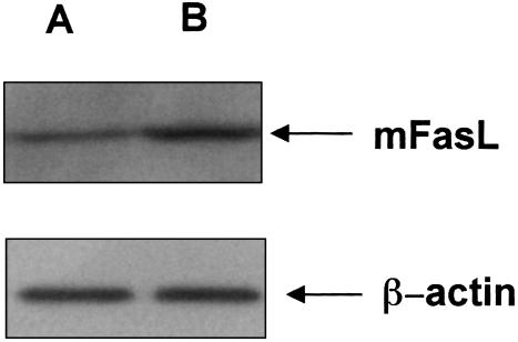
We examined in the present study the possible involvement of Fas and its ligand (FasL) in the process of Graves' disease. Immunohistochemical analysis showed that few normal thyrocytes expressed Fas but many thyrocytes in Graves' disease expressed this molecule. The percentage of FasL-positive thyrocytes in Graves' thyroids was, however, less than in normal thyroids. Several apoptotic thyrocytes and infiltrating mononuclear cells (MNCs) were detected scattered throughout Graves' thyroid tissues and abundant proliferating cell nuclear antigen (PCNA)-positive thyrocytes were present. Apoptotic cells, as well as PCNA-positive cells, were scarcely detectable in normal thyroid glands, however. In vitro treatment of thyrocytes by IL-1beta a cytokine found to be expressed in Graves' thyroid glands, increased Fas but reduced FasL expression. IL-1beta-stimulated thyrocytes became sensitive to apoptosis by anti-Fas IgM monoclonal antibody (mAb). Activated T cells, which strongly expressed FasL, showed cytotoxic activity toward IL-1beta-stimulated thyrocytes but not toward unstimulated thyrocytes. This cytotoxic activity involved the Fas/FasL pathway. Importantly, unstimulated thyrocytes could kill activated, but not resting, T cells. IL-1beta-stimulated thyrocytes, with down-regulated FasL expression, could not efficiently kill activated T cells. The cytotoxic activity of unstimulated thyrocytes toward activated T cells was inhibited by anti-FasL mAb. Interestingly, unstimulated thyrocytes induced apoptosis in IL-1beta-stimulated thyrocytes but not in unstimulated thyrocytes. These interactions were also blocked by anti-FasL mAb. Our results suggest that the apoptotic cell death of both thyrocytes and infiltrating MNCs found in Graves' thyroid glands is regulated by IL-1beta through Fas/FasL interactions.


Figures

Fig. 1
Fig. 1
Representative sections showing Fas and FasL expression in thyroid tissues from (a, c) normal thyroid and (b, d) Graves' disease. (a, b) Immunohistochemical staining with anti-Fas polyclonal antibodies. Magnification, ×400. (c, d) Immunohistochemical staining using anti-FasL polyclonal antibodies. Magnification, ×400.
Fig. 2
Fig. 2
Detection of in situ DNA breaks by TUNEL in thyroid tissues from (a) normal thyroid and (b–d) Graves' disease. Open arrows indicate apoptotic nuclei of thyrocytes and closed arrows indicate those of infiltrating mononuclear cells. Magnification, ×400.
Fig. 3
Fig. 3
Immunohistochemical staining of thyroid tissues (a) from normal thyroid and (b) Graves' disease using anti-PCNA mAb. Magnification, ×400.
Fig. 4
Fig. 4
IL-1β expression in thyroid tissues and effects of IL-1β on Fas and FasL expression on thyrocytes. (a, b) Immunohistochemical staining using IL-1β mAb. Magnification, × 400. (c) Flow cytometric analysis of Fas expression on cultured thyrocytes. Cells were labelled with control IgG (grey) or anti Fas mAb (black). Thyrocytes from Graves' thyroid tissues were stimulated with or without IL-1β (20 IU/ml) for 48 h. Upper: unstimulated thyrocytes, lower: IL-1β-stimulated thyrocytes. (d, e) DNA fragmentation of unstimulated or IL-1β-stimulated thyrocytes induced by anti-Fas IgM. Thyrocytes were cultured in the presence or absence of IL-1β, then treated with anti-Fas IgM for 18 h. DNA fragmentation was detected by Hoechst 33258 dye staining. (d) unstimulated thyrocytes (e) IL-1β-stimulated thyrocytes. Magnification, ×800. (f) Effects of IL-1β on FasL expression of thyrocytes. Thyrocytes from patients with Graves' disease were stimulated with various amounts of IL-1β for 0–72 h. Cells were lysed and the lysates were analysed by immunoblot using anti-FasL mAb. Left: dose-dependency studies, Right: time course studies (IL-1β: 20 IU/ml). Data shown (Fig. 2a–f) are representative experiment from five experiments.
Fig. 5
Fig. 5
FasL expression on resting or activated T cells from normal subjects. mFasL expression in resting (A) or activated (B) T cells was examined by Western blot analysis as described in the text. Representative data one experiment are shown. The experiment was repeated four times.
Fig. 6
Fig. 6
Cytotoxicity of T cells toward unstimulated or IL-1β-stimulated thyrocytes. (a) Effector cells are resting or activated T cells. Target cells are 51Cr-labelled unstimulated or IL-1β-stimulated thyrocytes. Effector cell to Target cell ratio was 20:1. A mixture of both cell types was incubated for 6 h. Cytotoxic activity was determined by 51Cr-release assay. Values are mean SD of percentages of cytotoxic activity of four experiments. (b) Blocking of cytotoxic activity of activated T cells toward unstimulated or IL-1β-stimulated thyrocytes by anti-FasL mAb. Activated T cells were pretreated with or without anti-FasL mAb, and then cocultured with 51Cr-labelled unstimulated or IL-1β-stimulated thyrocytes. Left: cytotoxicity toward unstimulated thyrocytes, Right: cytotoxicity toward IL-1β-stimulated thyrocytes. Anti-FasL mAb (–): Addition of control hamster IgG instead of anti-FasL mAb. Values are mean SD of percentages of cytotoxic activity of four experiments.
Fig. 7
Fig. 7
Functional Fas expression on activated T cells. (a, b) A representative example of the flow cytometric profile of Fas expression on T cells. Peripheral blood T cells were cultured with IL-2 for 7 days then incubated with PMA and ionomycin for 24 h. T cells were then incubated with either control mouse IgG (solid) or anti-Fas mAb (open). (a) Fas expression on resting T cells, (b) Fas expression on activated T cells. (c) DNA ladder formation in resting and activated T cells. DNA was extracted from anti-Fas IgM mAb-treated resting or activated T cells, and subjected to electrophoresis on a 1% agarose gel to detect the nucleosomal DNA ladder. DNA ladder formation was not detected in resting (Lane A) but only in activated T cells (Lane B) treated with anti-Fas IgM mAb. Lane C is a DNA size marker. Representative results are shown (Fig. 5a–c). The experiment was performed five times.
Fig. 8
Fig. 8
Cytotoxicity of thyrocytes toward resting or activated T cells. (a) Effector cells are unstimulated thyrocytes, and target cells are 51Cr-labelled resting or activated T cells. The effector cell to target cell ratio was 1 : 10. A mixture of effector and target cells was incubated for 6 h and cytotoxic activity was determined by 51Cr release assay. Values are mean SD of percentages of cytotoxic activity of four experiments. (b) Blocking of cytotoxic activity of thyrocytes against activated T cells by anti-FasL mAb. Thyrocytes were pretreated with or without anti-FasL mAb, and then cultured with 51Cr-labelled activated T cells. Anti-FasL mAb (–): Addition of control hamster IgG instead of anti-FasL mAb. Values are mean SD of percentages of cytotoxic activity of four experiments.
Fig. 9
Fig. 9
Fas/FasL interaction between unstimulated thyrocytes and IL-1β-stimulated thyrocytes. Thyrocytes separated from thyroid tissues of patients with Graves' disease were cultured with or without 20 IU/ml of IL-1β for 48 h. IL-1β-stimulated thyrocytes were labelled with 51Cr for 2 h, and 51Cr -labelled IL-1β-stimulated thyrocytes were used as target cells. Unstimulated or IL-1β-stimulated thyrocytes were used as effector cells. The effector to target cell ratio was 1:1. In blocking experiments, effector cells were pretreated with anti-FasL mAb for 30 min and then cocultured with 51Cr-labelled target cells. Ordinate: cytotoxic activity (%) of effector cells toward target cells. Effector cells (–): Unstimlated thyrocytes used for effector cells. Anti-FasL mAb (–): Addition of control hamster IgG instead of anti-FasL mAb. Values are mean SD of percentages of specific 51Cr release from target cells of four experiments.

References

    1. Nagata S, Golstein P. The Fas death factor. Science. 1995;267:1449–55. - PubMed
    1. Green DR. Apoptotic pathways: The roads to ruin. Cell. 1998;94:695–8. - PubMed
    1. Raff M. Cell suicide for beginners. Nature. 1998;396:119–22. - PubMed
    1. Dayan CM, Daniels GH. Chronic autoimmune thyroiditis. N Engl J Med. 1996;335:99–107. - PubMed
    1. Weetman AP, McGregor AM. Autoimmune thyroid disease: Further developments in our understanding. Endocr Rev. 1994;15:788–830. - PubMed

Publication types

MeSH terms