. 2001 Jul;35(1):26-34.
doi: 10.1002/glia.1067.

Transplantation of an acutely isolated bone marrow fraction repairs demyelinated adult rat spinal cord axons

Affiliations

Transplantation of an acutely isolated bone marrow fraction repairs demyelinated adult rat spinal cord axons

M Sasaki et al. Glia. 2001 Jul.

Abstract

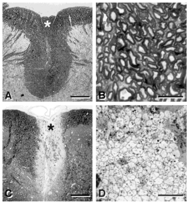
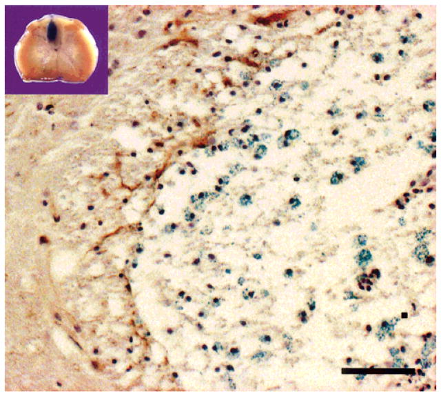
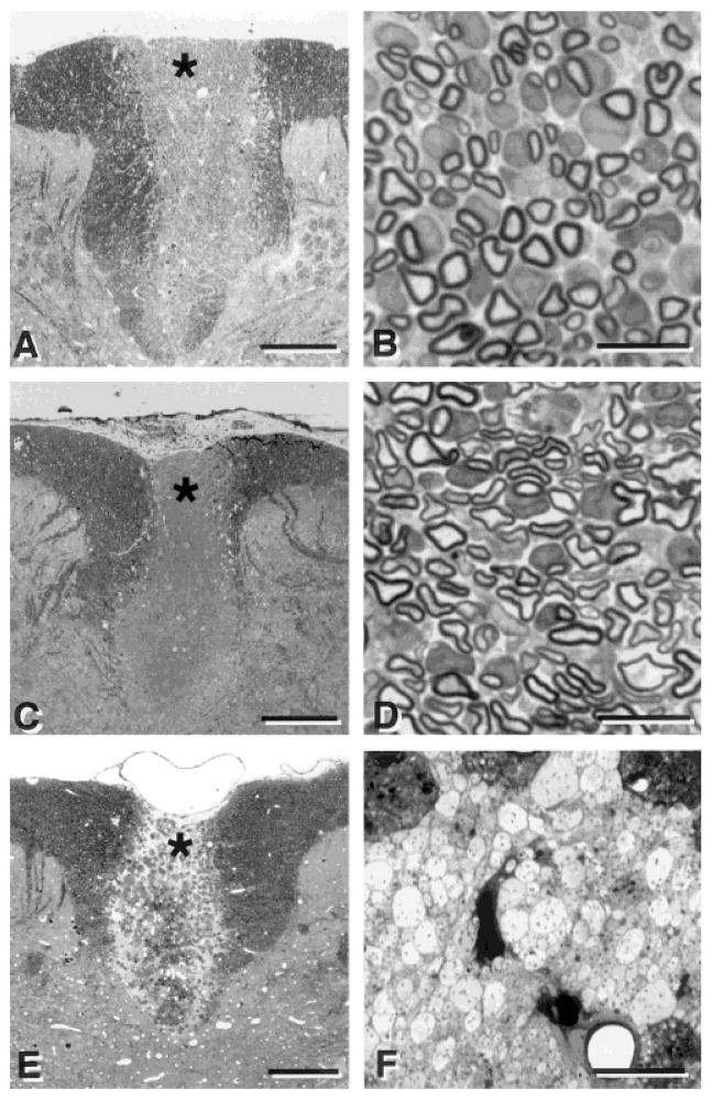
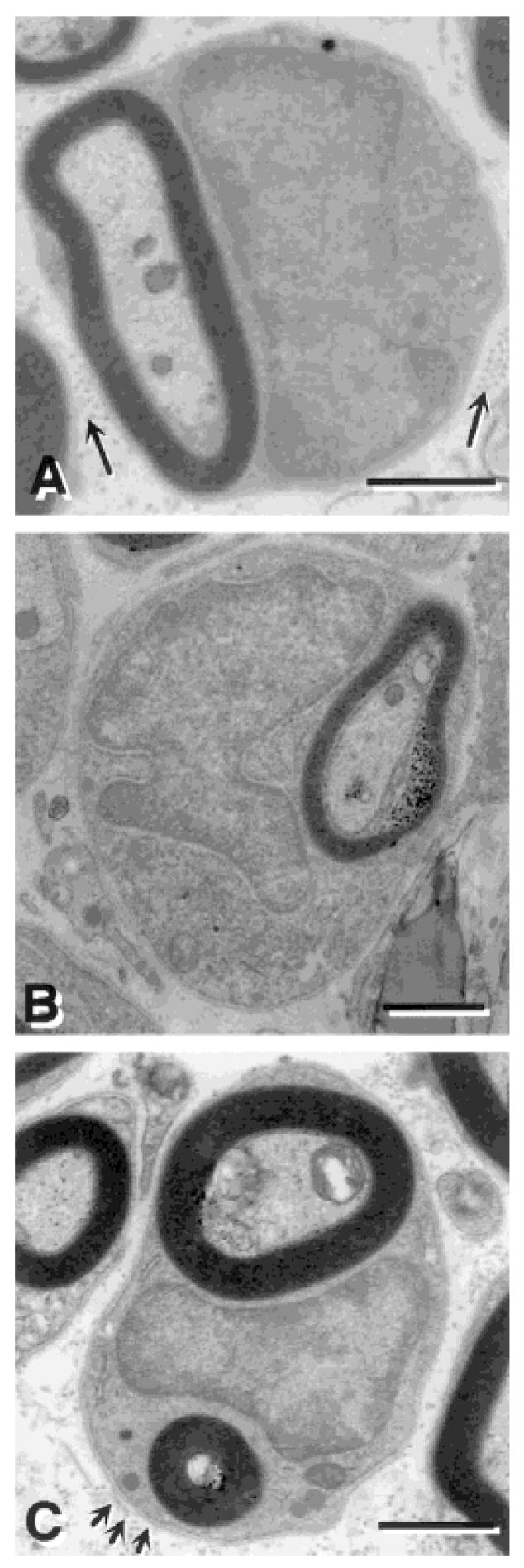
The potential of bone marrow cells to differentiate into myelin-forming cells and to repair the demyelinated rat spinal cord in vivo was studied using cell transplantation techniques. The dorsal funiculus of the spinal cord was demyelinated by x-irradiation treatment, followed by microinjection of ethidium bromide. Suspensions of a bone marrow cell fraction acutely isolated from femoral bones in LacZ transgenic mice were prepared by centrifugation on a density gradient (Ficoll-Paque) to remove erythrocytes, platelets, and debris. The isolated cell fraction contained hematopoietic and nonhematopoietic stem and precursor cells and lymphocytes. The cells were transplanted into the demyelinated dorsal column lesions of immunosuppressed rats. An intense blue beta-galactosidase reaction was observed in the transplantation zone. The genetically labeled bone marrow cells remyelinated the spinal cord with predominately a peripheral pattern of myelination reminiscent of Schwann cell myelination. Transplantation of CD34(+) hematopoietic stem cells survived in the lesion, but did not form myelin. These results indicate that bone marrow cells can differentiate in vivo into myelin-forming cells and repair demyelinated CNS.


Figures

Fig. 1
Fig. 1
Light micrographs of transverse sections of the dorsal spinal cord showing the dorsal funiculus in normal (A) and demyelinated (C) rats. Examination at higher magnification shows normal (B) and demyelinated (D) axons in the dorsal columns. B and D were prepared from the area around the asterisk in A and C, respectively. Scale bar = 250 μm in A,C; 10 μm in B,D.
Fig. 2
Fig. 2
Inset shows intense blue β-galactosidase reaction product after transplantation of LacZ transgenic mouse bone marrow cells into the central region of a demyelinated lesion in the rat spinal cord. Numerous cells with β-galactosidase blue reaction are observed in the demyelinated dorsal funiculus 3 weeks after transplantation of LacZ transgenic mouse bone marrow cells. The image is from the upper left region of the dorsal funiculus at the border of the lesion with lateral white matter. The section was processed for β-galactosidase and GFAP and counterstained with hematoxylin to observe nuclei. Scale bar = 25 μm.
Fig. 3
Fig. 3
Low- (left) and high- (right) power micrographs of the dorsal funiculus after transplantation of bone marrow cells (A,B), Schwann cells (C,D) and CD34+ hematopoietic cells (E,F). Note the relatively extensive myelination after transplantation of bone marrow cells (B) and Schwann cells (D). Transplantation of CD34+ hematopoietic cells derived from mouse bone marrow into the demyelinated rat spinal cord did not result in myelination, although the transplanted cells were present and survived in the demyelinated lesion. B, D, and F are prepared from the area around the asterisk in A, C and E, respectively. Scale bar = 250 μm in A,C,E; 10 μm in B,D,F.
Fig. 4
Fig. 4
Electron micrographs of remyelinated axons in the dorsal funiculus of the spinal cord after bone marrow transplantation. A: Many myelin-forming cells were similar to peripheral myelin-forming cells, characterized by large nuclear and cytoplasmic regions, and collagen (arrows) in the extracellular space. B: Several of the myelin-forming cells have multilobular nuclei in the BM transplantation group. C: Some of the myelin-forming cells in the BM transplantation group engaged more than one axon, while no Schwann cells were observed to engage more than one axon. Arrows in C point to a basement membrane surrounding the myelin-forming cell. Scale bar = 1 μm in A,B; 2 μm in C.
Fig. 5
Fig. 5
Comparison of axon diameter between bone marrow (BM) transplant group and Schwann cell (SC) transplant group. Nucleus (A) and cell body (B) sizes are plotted versus axon diameters.

References

    1. Akiyama Y, Honmou O, Kato T, Uede T, Hashi K, Kocsis JD. Transplantation of clonal neural precursor cells derived from adult human brain establishes functional peripheral myelin in the rat spinal cord. Exp Neurol. 2001;167:27–39. - PubMed
    1. Archer DR, Leven S, Duncan ID. Myelination by cryopreserved xenografts and allografts in the myelin-deficient rat. Exp Neurol. 1994;125:268–277. - PubMed
    1. Azizi SA, Stokes D, Augelli BJ, DiGirolamo C, Prockop DJ. Engraftment and migration of human bone marrow stromal cells implanted in the brains of albino rats—similarities to astrocyte grafts. Proc Natl Acad Sci USA. 1998;95:3908–3913. - PMC - PubMed
    1. Berthold C, Rydmark M. Morphology of normal peripheral axons. In: Waxman SG, Kocsis JD, Stys PK, editors. The axon: structure, function and pathophysiology. New York: Oxford University Press; 1995. pp. 13–50.
    1. Bjornson CR, Rietze RL, Reynolds BA, Magli MC, Vescovi AL. Turning brain into blood: a hematopoietic fate adopted by adult neural stem cells in vivo. Science. 1999;283:534–537. - PubMed

Publication types

MeSH terms