. 2001 Jul;67(7):2922-6.
doi: 10.1128/AEM.67.7.2922-2926.2001.

Photosynthetic and phylogenetic primers for detection of anoxygenic phototrophs in natural environments

Affiliations

Photosynthetic and phylogenetic primers for detection of anoxygenic phototrophs in natural environments

L A Achenbach et al. Appl Environ Microbiol. 2001 Jul.

Abstract

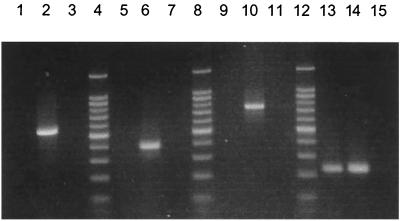
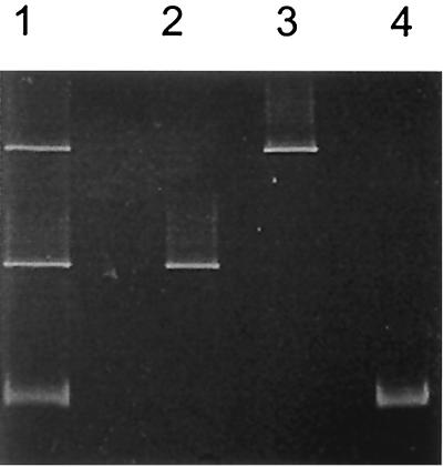
Primer sets were designed to target specific 16S ribosomal DNA (rDNA) sequences of photosynthetic bacteria, including the green sulfur bacteria, the green nonsulfur bacteria, and the members of the Heliobacteriaceae (a gram-positive phylum). Due to the phylogenetic diversity of purple sulfur and purple nonsulfur phototrophs, the 16S rDNA gene was not an appropriate target for phylogenetic rDNA primers. Thus, a primer set was designed that targets the pufM gene, encoding the M subunit of the photosynthetic reaction center, which is universally distributed among purple phototrophic bacteria. The pufM primer set amplified DNAs not only from purple sulfur and purple nonsulfur phototrophs but also from Chloroflexus species, which also produce a reaction center like that of the purple bacteria. Although the purple bacterial reaction center structurally resembles green plant photosystem II, the pufM primers did not amplify cyanobacterial DNA, further indicating their specificity for purple anoxyphototrophs. This combination of phylogenetic- and photosynthesis-specific primers covers all groups of known anoxygenic phototrophs and as such shows promise as a molecular tool for the rapid assessment of natural samples in ecological studies of these organisms.


Figures

FIG. 1
FIG. 1
Agarose gel of amplification products obtained using the phototroph-specific primer sets. LF 10 m is DNA from an environmental sample of the water column of Lake Fryxell, Antarctica, collected at a depth of 10 m; only the pufM primer set resulted in a positive amplification using this sample (lane 13). The negative controls contained no DNA template. Lanes 4, 8, and 12, 100-bp molecular size marker; lanes 1 to 3, green sulfur primer set (LF 10 m, Chlorobium tepidum, and negative control, respectively); lanes 5 to 7, green nonsulfur primer set (LF 10 m, Chloroflexus aurantiacus, and negative control, respectively); lanes 9 to 11, heliobacterial primer set (LF 10 m, Heliorestis daurensis, and negative control, respectively); lanes 13 to 15, pufM primer set (LF 10 m, Rhodocyclus purpureus, and negative control, respectively).
FIG. 2
FIG. 2
Agarose gels of amplification products from environmental samples using the specific primer sets. (A) PCR using the green sulfur bacterial primer set (amplified fragments are 525 bp). Lane 1, Pendant Lake, Antarctica; lane 2, New Zealand hot spring JP-1; lane 3, 1-kb ladder. (B) PCR using the green nonsulfur bacterial primer set (amplified fragments are 384 bp). Lane 1, Yellowstone hot spring New Pit; lane 2, Yellowstone hot spring Bath Lake; lane 3, 1-kb ladder. (C) PCR using the pufM primer set (amplified fragments are 229 bp). Lane 1, Yellowstone hot spring New Pit; lane 2, Lake Fryxell, Antarctica; lane 3, 100-bp ladder.
FIG. 3
FIG. 3
DGGE gel of pufM amplification products from Rhodobacter sphaeroides (lane 2), Rhodobacter palustris (lane 3), Thermochromatium tepidum (lane 4), and a mixture of all three bacterial species (lane 1).

References

    1. Amann R I, Ludwig W, Schileifer K. Phylogenetic identification and in situ detection of individual microbial cells without cultivation. Microbiol Rev. 1995;59:143–169. - PMC - PubMed
    1. Benson D A, Karsch-Mizrachi I, Lipman D J, Ostell J, Rapp B A, Wheeler D L. GenBank. Nucleic Acids Res. 2000;28:8–15. - PMC - PubMed
    1. Bryantseva I A, Gorlenko V M, Kompantseva E I, Achenbach L A, Madigan M T. Heliorestis daurensis gen. nov. sp. nov., an alkaliphilic coiled to rod-shaped phototrophic heliobacterium from an alkaline Siberian soda lake. Arch Microbiol. 1999;172:167–174. - PubMed
    1. Cahill A D, Stolz J F. Polyclonal antibodies to chlorosome proteins as probes for green sulfur bacteria. Appl Environ Microbiol. 1995;61:784–787. - PMC - PubMed
    1. Casamayor E O, Calderón-Paz J I, Mas J, Pedrós-Alió C. Identification of phototrophic sulfur bacteria through the analysis of lmwRNA band patterns. Arch Microbiol. 1998;170:269–278. - PubMed

Publication types

LinkOut - more resources