. 2001 Jul 1;21(13):4637-48.
doi: 10.1523/JNEUROSCI.21-13-04637.2001.

The OMP-lacZ transgene mimics the unusual expression pattern of OR-Z6, a new odorant receptor gene on mouse chromosome 6: implication for locus-dependent gene expression

Affiliations

The OMP-lacZ transgene mimics the unusual expression pattern of OR-Z6, a new odorant receptor gene on mouse chromosome 6: implication for locus-dependent gene expression

M Pyrski et al. J Neurosci. .

Erratum in

  • J Neurosci 2001 Aug 15;21(16):6457

Abstract

Reporter gene expression in the olfactory epithelium of H-lacZ6 transgenic mice mimics the cell-selective expression pattern known for some odorant receptor genes. The transgene construct in these mice consists of the lacZ coding region, driven by the proximal olfactory marker protein (OMP) gene promoter, and shows expression in a zonally confined subpopulation of olfactory neurons. To address mechanisms underlying the odorant receptor-like expression pattern of the lacZ construct, we analyzed the transgene-flanking region and identified OR-Z6, the first cloned odorant receptor gene that maps to mouse chromosome 6. OR-Z6 bears the highest sequence similarity (85%) to a human odorant receptor gene at the syntenic location on human chromosome 7. We analyzed the expression pattern of OR-Z6 in olfactory tissues of H-lacZ6 mice and show that it bears strong similarities to that mapped for beta-galactosidase. Expression of both genes in olfactory neurons is primarily restricted to the same medial subregion of the olfactory epithelium. Axons from both neuronal subpopulations project to the same ventromedial aspect of the anterior olfactory bulbs. Furthermore, colocalization analyses in H-lacZ6 mice demonstrate that OR-Z6-reactive glomeruli receive axonal input from lacZ-positive neurons as well. These results suggest that the expression of both genes is coordinated and that transgene expression in H-lacZ6 mice is regulated by locus-dependent mechanisms.


Figures
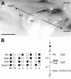
Fig. 1.
Fig. 1.
Expression pattern and chromosomal location of theOMPlacZ transgene in H-lacZ6 mice. A, X-gal staining in the right heminose of a 3-week-old H-lacZ6 mouse is shown. The punctate pattern shown corresponds to the ORN subpopulation expressing the lacZ transgene (black dots). The majority of β-gal+ neurons is concentrated on endoturbinate-IIv(star); fewer neurons are scattered along the rostrocaudal axis of the OE (arrows).Endoturbinate-IId and -IIv, Dorsal and ventral aspects of endoturbinate-II. Scale bar, 1 mm. B, The transgene insertion site in H-lacZ6 mice (Hlz6 locus) maps to the proximal region of mouse chromosome 6, as determined by interspecific backcross analysis. Left, The segregation patterns ofHlz6 and the flanking genes (Ptn,Tcrb, and Hoxa5) were typed in 174 backcross animals. For individual pairs of loci, >174 animals were typed (see text). Each column represents the chromosome identified in the backcross progeny that was inherited from the C57BL/6J × M. spretus F1 parent. Theblack and white boxes refer to the presence of a C57BL/6J allele and an M. spretus allele, respectively. The number of offspring inheriting each type of chromosome is listed at the bottom of eachcolumn. Right, A partial chromosome 6 linkage map shows the location of Hlz6 in relation to its linked genes. Recombination distances between loci in centimorgans are shown to the left of the chromosome, and the positions of loci in human chromosomes, where known, are shown to theright. References for the human map positions of loci cited in this study can be obtained from the Genome Data Base, a computerized database of human linkage information maintained by The William H. Welch Medical Library of The Johns Hopkins University (Baltimore, MD).
Fig. 2.
Fig. 2.
Identification of the new odorant receptor geneOR-Z6 at the Hlz6 locus.A, Gel electrophoresis is shown of the products obtained after PCR of the four P1 clones 6386–6389 (lanes 5–8) using the two different primer sets primers p26 and p27 and primers ps2 and ps4. All P1 clones carry the 3′-flanking fragment as demonstrated by the 160 bp amplicon derived from primers ps2 and ps4 (lanes 5–8). The 390 bp PCR product (star) derived from the degenerate OR primers p26 and p27 is only evident for P1 clone 6386 (lane 5) and the positive control (+C, lane 4) using mouse genomic DNA. The thickness of the band in the positive control (lane 4) reflects the amplification of numerous genomic OR genes. The positive control for primers ps2 and ps4 (+C, lane 2; mouse genomic DNA) shows a 160 bp amplicon, whereas negative control reactions, omitting template DNA, yielded no products for primers ps2 and ps4 (−C,lane 1) or primers p26 and p27 (−C,lane 3). HaeIII-digested ΦX174 phage DNA (M) is the length standard (lane 9). Fragment sizes in base pairs are indicated by thenumbers at the right.B, Restriction digestion (left) followed by Southern blot hybridization (right) shows that the P1 clone 6386 carries a single OR gene. Eight hundred nanograms of each P1 6386 DNA digest (E, EcoRI;S, SacI; X,XbaI) were resolved on a 0.6% agarose gel, transferred onto a nylon membrane, and subjected to Southern blot hybridization. The probe was prepared by labeling the 390 bp PCR fragment obtained inA with digoxigenin-conjugated dUTP via PCR using primers p26 and p27. Hybridizing fragment sizes were 4 kb (EcoRI), 11 kb (SacI), and 3 kb (XbaI). The 3 kb hybridizing XbaI fragment (arrowhead) was subcloned and sequenced.HindIII-digested λ DNA (M) is the length standard in kilobase pairs indicated by the numbers at theleft.
Fig. 3.
Fig. 3.
The new mouse OR-Z6 gene encodes a typical OR protein. A, Top, Partial restriction map of the 3 kb XbaI fragment carrying the complete OR-Z6 coding region (black box) and sequences 1.2 kb upstream and 1 kb downstream. Location of the gene-specific primers N and C used in RT-PCR analyses (C) is indicated by arrows. Restriction enzymes are the following: B,BamHI; E, EcoRI;S, SphI; X,XbaI. Bottom, Comparison of the deduced amino acid sequences of the coding regions of mouseOR-Z6 (mOR-Z6) and a humanOR-Z6 ortholog (hOR-Z6) inone-letter code. For hOR-Z6, only differences from the mOR-Z6 sequence are shown.Gray shading depicts the predicted seven transmembrane domains TM-1 to TM-7. The amino acid sequence domains of the two OR-Z6 proteins that match closely to the motifs conserved (indicated by a horizontal line) among OR proteins are PMYFFL (TM-2), MAVDRYVAVC (TM-3), SY (TM-5), K(A/S)FSTCASH (TM-6), and PFLNPF (TM-7). Both OR-Z6 proteins share 85% similarity over a total of 314 amino acids and exhibit cysteine residues in conserved positions (97, 179, stars) and a putative N-terminal receptor glycosylation site (arrowhead), as well as several serine and threonine phosphorylation sites in the third intracellular loop (IL-3). B, Genomic Southern blot hybridization for OR-Z6. Restriction digestion of mouse liver DNA (10 μg/lane) from wild-type (wt) and H-lacZ6 mice was as indicated (H,HindIII; S, SphI;X, XbaI). The Southern blot was hybridized with the digoxigenin-labeled OR-Z6 coding region. The DNA probe hybridized to a single fragment perlane, and the sizes of the hybridizing fragments for both wild-type and H-lacZ6 genomic DNA were identical (H, 23 kb; S, 1.72 kb; X, 3 kb). HindIII-digested λ DNA (M) is the length standard in kilobase pairs indicated by the numbers on theleft. C, RT-PCR performed with RNA from three different tissues demonstrating that OR-Z6 mRNA is expressed in olfactory tissues (OE, OB). The photograph shows a gel electrophoresis of the products derived from RT-PCR using the OR-Z6 specific primers N and C. The 899 bp amplicon was only evident in samples with preceding reverse transcription (+RT). No products were obtained from RT-PCR using liver mRNA (L) or from reactions omitting reverse transcriptase (−RT). The lower intensity of the fragment obtained from OB mRNA versus that from OE mRNA coincides with reports demonstrating that OR mRNA is much less abundant in axon terminals compared with their cell bodies (Ressler et al., 1994).
Fig. 4.
Fig. 4.
OR-Z6 mRNA and β-galactosidase exhibit similar expression patterns in mouse OE. The hemisections (left nostril) shown in both columns represent a survey from anterior (top) to posterior (bottom) in the mid-to-caudal region of the OE. Left column,In situ hybridization for OR-Z6(digoxigenin-labeled riboprobe) performed in an array of 15 μm coronal cryosections of the OE from a wild-type mouse (129S3; postnatal day 24) is shown. OR-Z6+ neurons are restricted to endoturbinate-II and -III and ectoturbinate-3 as indicated. Similar to lacZ+ neurons (right column), OR-Z6+neurons are located at different depths of the OE; the most intensely labeled neurons are located in the apical OE. Right column, The overall distribution of OR-Z6 mRNA is almost identical to the β-galactosidase expression pattern observed in an age-matched H-lacZ6 mouse. As seen forOR-Z6 (boxed area, left column), X-gal-stained ORNs (right column) are concentrated on the tips of the central turbinates. To facilitate the comparison of the expression patterns for OR-Z6 mRNA and β-galactosidase, sections were chosen to match similar regions of the OE. Scale bar, 500 μm.
Fig. 5.
Fig. 5.
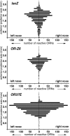
Anterior-to-posterior distribution of β-galactosidase (X-gal staining, top),OR-Z6 (in situ hybridization,middle), and OR37E (in situ hybridization, bottom) in separate but adjacent nose sections of the same P21 H-lacZ6 mouse. Starting at the organ of Masera (0 mm), 15 μm coronal cryosections were collected, and the labeled cells of every 10th section were counted and plotted along the anterior-to-posterior axis. The three expression profiles shown are nearly bilaterally symmetrical, and each of the three neuron phenotypes shows the highest density of reactive ORNs in the mid-to-caudal aspect of the OE at ∼2.6 mm posterior to the organ of Masera. ORNs expressing the lacZ transgene were approximately twice as numerous as wereOR-Z6+ neurons. Whereas a small number of β-gal+ ORNs was evident in the region up to 1.2 mm posterior to the organ of Masera, almost noOR-Z6+ or mOR37E+ neurons were found in this anterior aspect. The unusually large number of mOR37E+ neurons is caused by cross-hybridization. The riboprobe used derives from the mOR37E coding region that is highly homologous among the four members of this subfamily, all of which are expressed in the same zone (Strotmann et al., 2000). The total numbers of labeled ORNs in the left or right nose of the H-lacZ6 mouse analyzed were as follows: X-gal+, 5680 or 5780;OR-Z6+, 3400 or 3240; and mOR37E+, 10,230 or 10,410, respectively.
Fig. 6.
Fig. 6.
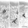
The three bright-field images illustrate the criteria by which olfactory neurons coexpressing thelacZ transgene and OR-Z6 were identified.OR-Z6 in situ hybridization was performed using a digoxigenin-labeled OR-Z6 riboprobe, and hybridization signals were detected and visualized using an alkaline phosphatase-conjugated anti-digoxigenin antibody and a substrate that developed a purple precipitate. Left, Hybridization signals were most prominent in the apical cytoplasmic portion and appeared triangular (arrows). In addition we observed a thin line surrounding the nucleus; the nucleus itself was usually white. Right, In contrast, ORNs that solely express the lacZ transgene (arrow) were gray, including the cytoplasm covering the nucleus. Expression of β-galactosidase was detected using a primary anti-β-galactosidase antibody and visualized using a substrate yielding a gray precipitate.Middle, The double-labeled neuron shows a combination of both features described. The gray cytoplasm covering the nucleus indicates β-galactosidase expression (arrow), whereas the peak-shaped purple precipitate in the apical cytoplasm corresponds to OR-Z6 in situ hybridization (arrowhead). The three differently labeled neurons shown derive from the same coronal cryosection of a 35-d-old H-lacZ6 mouse and were closely associated on endoturbinate-II. Scale bar, 20 μm.
Fig. 7.
Fig. 7.
OR-Z6 neurons preferentially project to a single glomerulus in the main OB. In situhybridization was performed on serial coronal cryosections of the entire OBs using a [32P]CTP-labeled antisense RNA probe of the OR-Z6 coding region. Subsequent counterstaining of periglomerular nuclei facilitated the identification of individual glomeruli in the glomerular layer (GL). Top panels, The dark-field photomicrographs illustrate that axonal projections ofOR-Z6-reactive neurons preferentially terminate onto a single ventromedial glomerulus in each OB, as depicted by thearrowheads. Bottom panels 1–3, The hybridization signals (white grains) shown for this specimen were evident in three consecutive 18 μm sections per bulb (boxed areas in top panels magnified).EPL, External plexiform layer; ONL, olfactory nerve layer. Scale bar: bottom panels 1–3, 50 μm.
Fig. 8.
Fig. 8.
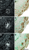
OR-Z6-positive glomeruli in H-lacZ6 mice receive input from two phenotypically different ORN populations. IHC for β-galactosidase and in situ hybridization for OR-Z6 were performed on separate but adjacent sets of coronal cryosections (15 μm) cut from the OB of a P26 H-lacZ6 mouse. The panelsshow a series of higher magnifications of the ventromedial portion of the left OB. The order in which sections were taken along the anterior-to-posterior axis is indicated by the numbers 1–6. Hybridization signals for OR-Z6 appear as dense white grains (dark-field, left), whereas β-gal+ axons are visible asbrown fibers entering their target glomeruli (bright-field, right). Corresponding glomeruli in adjacent sections were aligned after identifying glomerular borders by counterstaining periglomerular nuclei. Comparison of labeled glomeruli in the two columns demonstrates that theOR-Z6+ glomerulus receives input from β-gal+ axons as well (white boxed areas). The changing signal intensity for bothOR-Z6 and β-gal as the sections progress through the different depths of the glomerulus can be traced along the anterior-to-posterior axes of the three sections shown. Note the small number of β-gal+ axons projecting to theOR-Z6-labeled glomerulus. Scale bar, 100 μm.

References

    1. Asai H, Kasai H, Matsuda Y, Yamazaki N, Nagawa F, Sakano H, Tsuboi A. Genomic structure and transcription of a murine odorant receptor gene: differential initiation of transcription in the olfactory and testicular cells. Biochem Biophys Res Commun. 1996;221:240–247. - PubMed
    1. Astic L, Saucier D. Anatomical mapping of the neuroepithelial projection to the olfactory bulb in the rat. Brain Res Bull. 1986;16:445–454. - PubMed
    1. Ben-Arie N, Lancet D, Taylor C, Khen M, Walker N, Ledbetter DH, Carrozzo R, Patel K, Sheer D, Lehrach H. Olfactory receptor gene cluster on human chromosome 17: possible duplication of an ancestral receptor repertoire. Hum Mol Genet. 1994;3:229–235. - PubMed
    1. Buck L, Axel R. A novel multigene family may encode odorant receptors: a basis for odorant recognition. Cell. 1991;65:175–187. - PubMed
    1. Carver EA, Issel-Tarver L, Rine J, Olsen AS, Stubbs L. Location of mouse and human genes corresponding to conserved canine olfactory receptor gene family. Mamm Genome. 1998;9:349–354. - PubMed

Publication types

MeSH terms

Associated data