. 2001 Jul 1;21(13):4668-77.
doi: 10.1523/JNEUROSCI.21-13-04668.2001.

Activation of the nuclear factor-kappaB is a key event in brain tolerance

Affiliations

Activation of the nuclear factor-kappaB is a key event in brain tolerance

N Blondeau et al. J Neurosci. .

Abstract

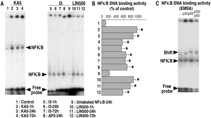
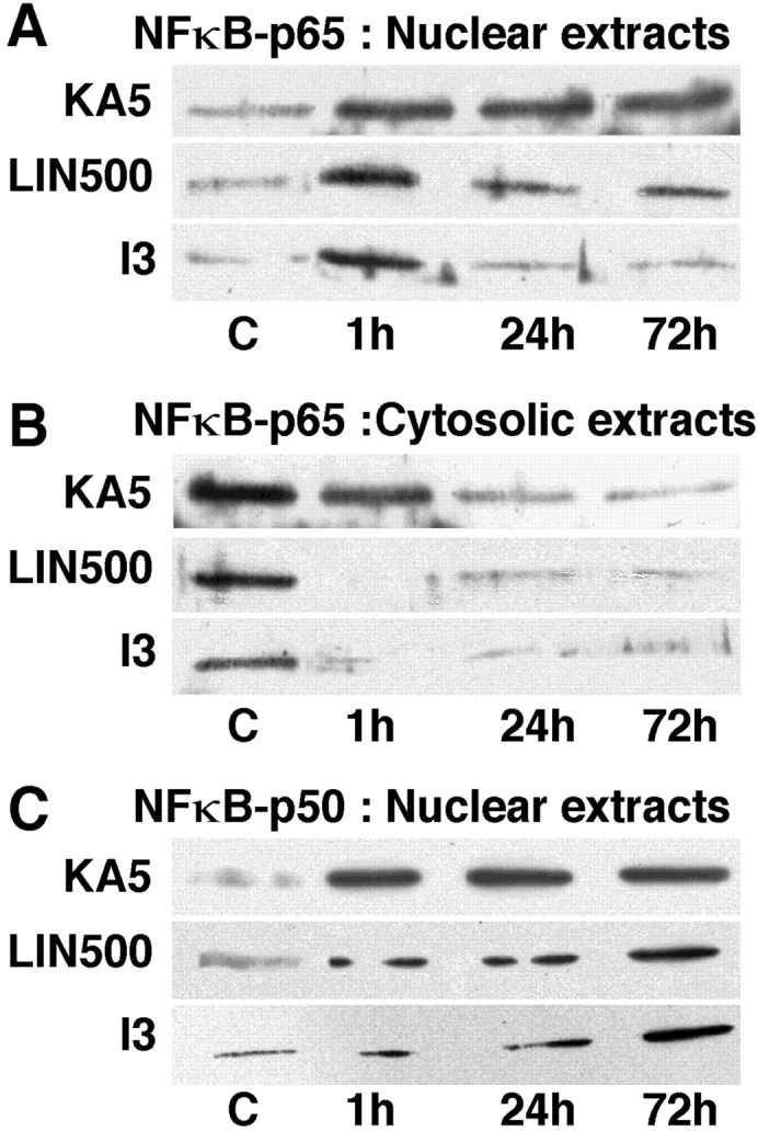
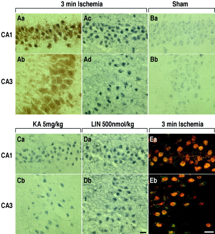
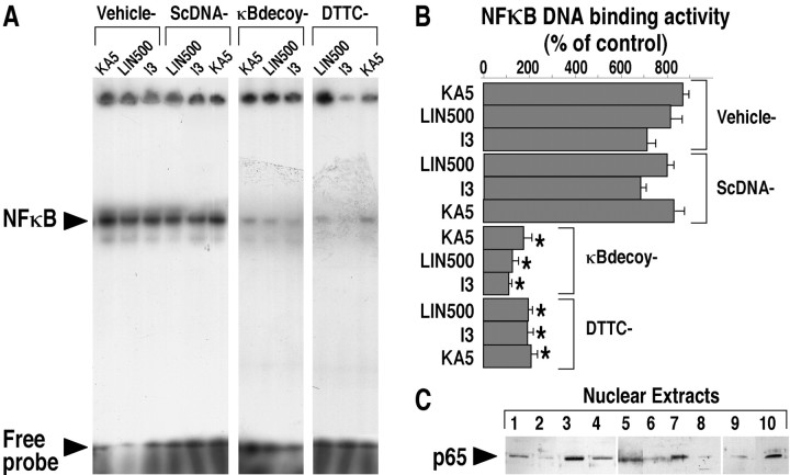
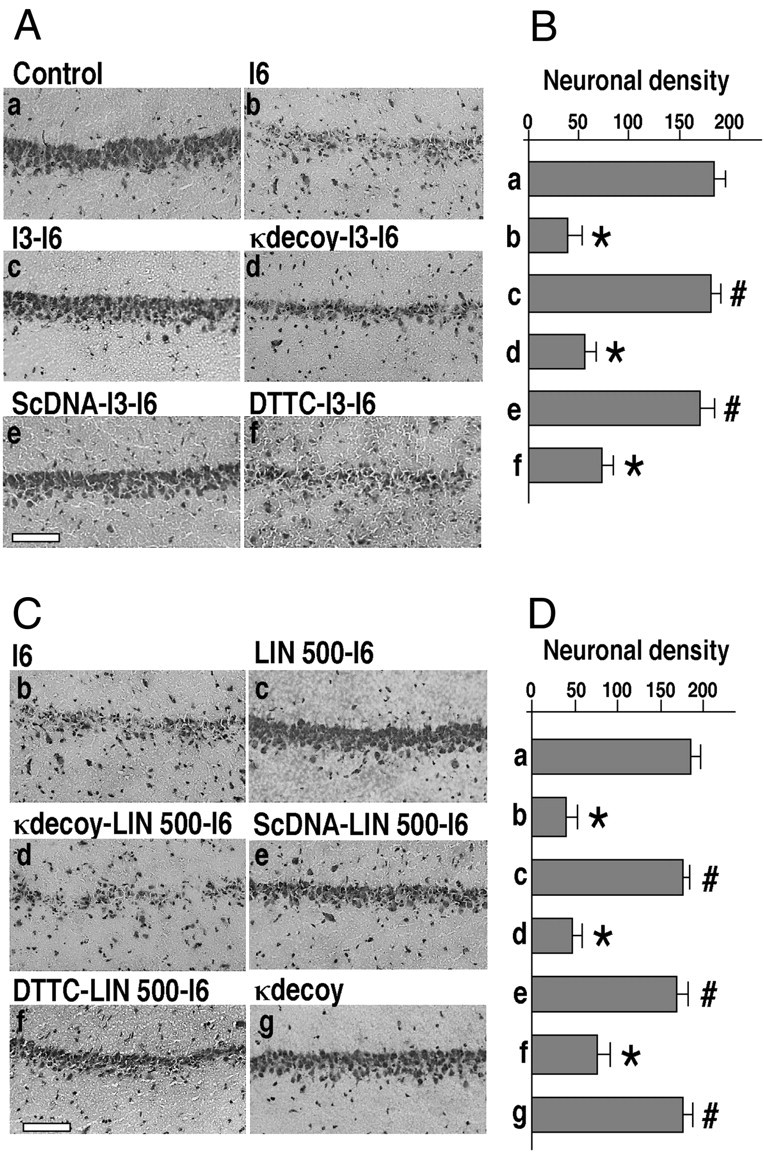
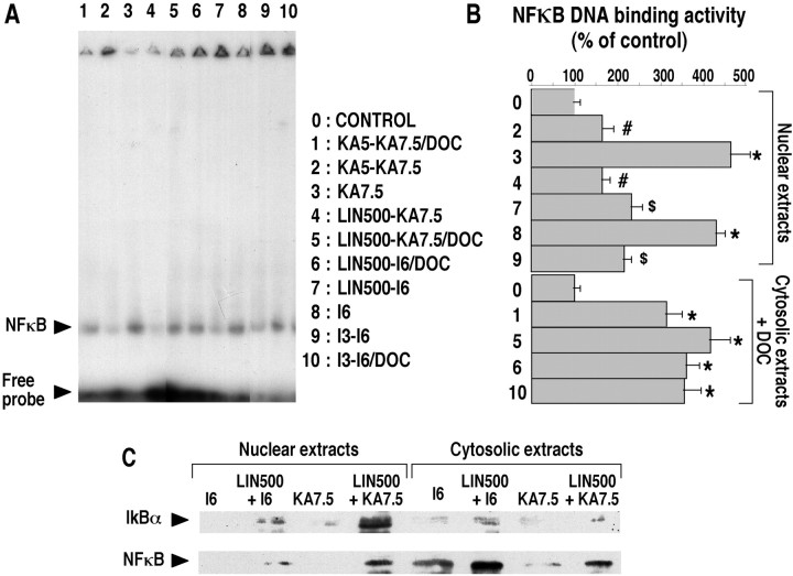
The transcription factor nuclear factor-kappaB (NFkappaB) is an ubiquitously expressed inducible regulator of a broad range of genes and plays a pivotal role in cell death and survival pathways. Three models of brain tolerance (ischemic, epileptic, and polyunsaturated fatty acid-induced preconditioning), known to confer resistance to neurons against ischemia or status epilepticus, were used to determine whether NFkappaB mediated the late preconditioning. A sublethal 3 min ischemia, a dose of 5 mg/kg kainic acid (KA5) or 500 nmol of linolenic acid (LIN500) led to a rapid increase of NFkappaB DNA-binding activity and nuclear translocation of p65 and p50 subunits of NFkappaB in neurons. Pretreatment with the NFkappaB inhibitor diethyldithiocarbamate or kappaB decoy DNA blocked the increased DNA-binding activity and the nuclear translocation of NFkappaB and abolished the neuroprotective effects of different delayed preconditionings against severe ischemia or epilepsy. The inhibition of NFkappaB observed in rats preconditioned with 3 min ischemia, KA5 or LIN500 treatments compared with ischemic or epileptic controls was correlated with the prevention of the inducible degradation of the inhibitory protein IkappaBalpha. Preconditioning probably inhibits the activation of NFkappaB by interfering with a pathway that leads to the direct transcriptional activation of IkappaBalpha by NFkappaB itself. The present work provides evidence that activation of NFkappaB is a crucial step in the signal transduction pathway that underlies the development of brain tolerance and may open new strategies in the prevention of cerebral diseases, such as ischemia or epilepsy.


Figures

Fig. 1.
Fig. 1.
EMSA showing the time course of increased NFκB DNA-binding activity induced by the three different preconditionings (3 min ischemia, KA5 treatment, and LIN500 treatment).A, Representative gel shifts analysis showing NFκB DNA-binding activity in nuclear protein extracts from hippocampi of control rats (lane 1) or rats submitted to 3 min ischemia (lanes 5–9) or treated with KA5 (lanes 2–4) or LIN500 (lanes 10–12) and obtained at 1, 24, and 72 hr after the different preconditionings. NFκB DNA-binding activity was assayed as described in Materials and Methods. Competition assays of NFκB DNA-binding activity was performed in the presence of 50-fold excess of unlabeled competitor NFκB (lane 9) and nonspecific competitor AP2 (lane 8) consensus oligonucleotides. The shifted bands of specific NFκB DNA complexes are indicated by thearrowheads. The right andleft gels correspond to 4 and 7% polyacrylamide gels, respectively. B, Quantification of NFκB DNA-binding activity in the different experimental groups. The specific shifted bands were quantified using a phosphorimaging system as described in Materials and Methods. The values are expressed as a percentage of control. No significant differences were found between vehicle-injected and sham-operated rats, and values of these groups were pooled and termed control. Data represent the mean ± SEM values. Data are representative of six separate experiments in each group (n = 6). *p < 0.05 indicates statistical significance when compared with control.C, Supershift analysis of NFκB binding proteins present in nuclear extracts 24 hr after 3 min ischemia. The binding activity assay was performed in the presence of anti-p65 antibodies, anti-p50 antibodies, a combination of both, or no antibody. Thearrowheads indicate the bands of specific NFκB–DNA complexes, which were supershifted by anti-p65 antibodies, anti-p50 antibodies, or by their combination.
Fig. 2.
Fig. 2.
Representative Western blotting analysis of p65 and p50 subunits of NFκB in nuclear (A,C) and cytosolic (B) extracts from hippocampi of control rats (C) or rats submitted to 3 min ischemia (I3) or treated with KA5 or LIN500 and obtained at 1, 24, and 72 hr after the different preconditionings.
Fig. 3.
Fig. 3.
Representative immunohistochemical staining of the active form of NFκB protein in hippocampal CA1 and CA3 substructures of rats killed 24 hr after each of preconditioning stimuli.Aa–Ad, 3 min ischemia. Ba,Bb, Sham-surgery. Ca, Cb, KA5 treatment. Da, Db, LIN500 treatment.Aa, Ab, Immunohistochemical staining of NeuN protein within CA1 and CA3 pyramidal cells after sublethal ischemia. E, Colocalization of NFκB-p65 and the neuron-specific nuclear protein NeuN (neuronal nuclei) within CA1 (Ea) and CA3 (Eb) pyramidal cells. Brain sections were immunostained with antibodies against the p65 subunit of NFκB (green) and NeuN (red) for double-labeling. Scale bars, 20 μm.
Fig. 4.
Fig. 4.
Electrophoretic mobility shift assay (A, B) and Western blotting analysis of p65 subunit of NFκB (C) showing the effect of intracerebroventricular administration of κB decoy DNA or intraperitoneal injection of DTTC on the increased NFκB DNA-binding activity induced by the three different preconditionings (3 min ischemia, KA5 treatment, and LIN500 treatments). A, Representative gel shift analysis showing NFκB DNA-binding activity in hippocampal nuclear extracts isolated from rats that had received intracerebroventricular injections of vehicle, 60 μg of κB decoy DNA (κBdecoy), 60 μg of scrambled control DNA (ScDNA), or intraperitoneal injection of DTTC (150 mg/kg) before sublethal ischemia (I3) or administration of either KA5 or LIN500. Rats were killed 1 hr (for κB decoy or scrambled control DNA) or 24 hr (for DTTC) after each type of preconditioning. NFκB activity was assayed as described in Materials and Methods. The shifted band of specific NFκB DNA complexes is indicated by the arrowhead. B, Quantification of NFκB DNA-binding activity in the different experimental groups. The specific shifted bands were quantified using a phosphorimaging system as described in Materials and Methods. Values are expressed as a percentage of control. Results are expressed as mean ± SEM. Data are representative of six separate experiments in each group (n = 6). *p < 0.05 indicates statistical significance when compared with vehicle-injected rats. C, Representative Western blotting analysis of p65 subunit of NFκB in hippocampal nuclear extracts isolated from rats that had received intracerebroventricular injections of vehicle, 60 μg of κB decoy DNA (κBdecoy), 60 μg of scrambled control DNA (ScDNA) or intraperitoneal DTTC injection (150 mg/kg) before I3 or LIN500. Rats were killed 24 hr after the conditioning stimulus. The position of p65 is indicated by thearrowhead. 1, Vehicle; 2, DTTC–LIN500; 3, LIN500; 4, Sham-operated; 5, I3; 6, DTTC–I3;7, scrambled control DNA–LIN500; 8, κB decoy DNA–LIN500; 9, κB decoy DNA–I3;10, scrambled control DNA–I3.
Fig. 5.
Fig. 5.
Effect of κB decoy DNA, scrambled control DNA, and DTTC injections on the ischemic tolerance induced by brief ischemia or LIN500 treatment. A, C, Representative photomicrographs highlighting morphological changes in CA1 subfield of cresyl violet-stained hippocampal sections 7 d after severe 6 min ischemia in the different experimental groups. I6corresponds to rats submitted to 6 min ischemia. I3-I6corresponds to rats submitted to 3 min ischemia 3 d before 6 min ischemia. κdecoy I3-I6 or ScDNA I3-I6corresponds to rats that, respectively, had received intracerebroventricular injections of 60 μg of κB decoy DNA or scrambled control DNA at 24 and 2 hr before 3 min ischemia. DTTC I3-I6 corresponds to rats that had received an intraperitoneal injection of DTTC (150 mg/kg) 30 min before 3 min ischemia. Rats were killed 7 d after 6 min ischemia. Scale bar, 100 μm.B, D, Quantification of neuronal density in the hippocampal CA1 pyramidal layer of different experimental groups. Results are expressed as mean ± SEM (n = 6) and represent neuronal densities assessed in cresyl violet-stained sections per 1 mm linear length of CA1 pyramidal layer. A mean value for each CA1 substructure was obtained from 10 bilateral measurements on two sections per slide and 10 slides per animal (n = 6) in each of the experimental groups. Differences were considered statistically significant whenp < 0.05 (Tukey's test). * indicates significantly different from control (sham-operated animals). # indicates significantly different from ischemic animals (6 min).
Fig. 6.
Fig. 6.
Changes in NFκB DNA-binding activity and subcellular localization of IκBα and active form of NFκB in the different groups of preconditioned brains. A, Representative gel shifts analysis showing NFκB DNA-binding activity in hippocampal nuclear and cytosolic extracts isolated from rats preconditioned with 3 min ischemia (I3-I6), KA5 treatment (KA5-KA7.5), or LIN500 treatment (LIN500-I6 or LIN500-KA7.5) and submitted to 6 min ischemia or KA7.5 treatment 3 d later. Rats were killed 24 hr after the last treatment. Cytosolic extracts were analyzed by EMSA with DOC treatment after binding as described in Materials and Methods. The shifted band of specific NFκB–DNA complexes is indicated by the arrowhead. B, Quantification of NFκB DNA-binding activity in the different experimental groups. The specific shifted bands were quantified using a phosphorimaging system as described in Materials and Methods. The values are expressed as a percentage of control. No significant differences were found between saline-injected and sham-operated rats, and values of these groups were pooled and termed control. Results are expressed as mean ± SEM. Data are representative of six separate experiments in each group (n = 6). Differences were considered statistically significant whenp < 0.05 (Tukey's test). * indicates significantly different from control (saline-injected or sham-operated animals). # indicates significantly different from KA7.5-injected animals. $ indicates significantly different from ischemic animals (6 min). C, Representative Western blotting analysis of IκBα and active form of NFκB in hippocampal nuclear and cytosolic extracts isolated from ischemic (I6) and epileptic (KA7.5) rats and rats preconditioned with LIN500 treatment (LIN500-I6 orLIN500-KA7.5) and submitted to severe 6 min ischemia or KA7.5 treatment 3 d later. Rats were killed 24 hr after the last treatment. The positions of IκBα and active form of NFκB are indicated by the arrowheads.

References

    1. Arenzana-Seisdedos F, Thompson J, Rodriguez MS, Bachelerie F, Thomas D, Hay RT. Inducible nuclear expression of newly synthesized I kappa B alpha negatively regulates DNA-binding and transcriptional activities of NF-kappa B. Mol Cell Biol. 1995;15:2689–2696. - PMC - PubMed
    1. Auphan N, DiDonato JA, Rosette C, Helmberg A, Karin M. Immunosuppression by glucocorticoids: inhibition of NF-kappa B activity through induction of I kappa B synthesis. Science. 1995;270:286–290. - PubMed
    1. Baeuerle PA, Baltimore D. NF-kappa B: ten years after. Cell. 1996;87:13–20. - PubMed
    1. Barger SW, Horster D, Furukawa K, Goodman Y, Krieglstein J, Mattson MP. Tumor necrosis factors alpha and beta protect neurons against amyloid beta-peptide toxicity: evidence for involvement of a kappa B-binding factor and attenuation of peroxide and Ca2+ accumulation. Proc Natl Acad Sci USA. 1995;92:9328–9332. - PMC - PubMed
    1. Blondeau N, Plamondon H, Richelme C, Heurteaux C, Lazdunski M. KATP channel openers, adenosine agonists and epileptic preconditioning are stress signals inducing hippocampal neuroprotection. Neuroscience. 2000;100:465–474. - PubMed

Publication types

MeSH terms