

Synaptic currents generating the inhibitory surround of ganglion cells in the mammalian retina
- PMID: 11425912
- PMCID: PMC6762364
- DOI: 10.1523/JNEUROSCI.21-13-04852.2001
Synaptic currents generating the inhibitory surround of ganglion cells in the mammalian retina
Abstract
The receptive field (RF) of retinal ganglion cells (RGCs) consists of an excitatory central region, the RF center, and an inhibitory peripheral region, the RF surround. It is still unknown in detail which inhibitory interneurons (horizontal or amacrine cells) and which inhibitory circuits (presynaptic or postsynaptic) generate the RF surround. To study surround inhibition, light-evoked whole-cell currents were recorded from RGCs of the isolated, intact rabbit retina. The RFs were stimulated with light or dark spots of increasing diameters and with annular light stimuli. Direct inhibitory currents could be isolated by voltage clamping ganglion cells close to the Na(+)/K(+) reversal potential. They mostly represent an input from GABAergic amacrine cells that contribute to the inhibitory surround of ganglion cells. This direct inhibitory input and its physiological function were also investigated by recording light-evoked action potentials of RGCs in the current-clamp mode and by changing the intracellular Cl(-) concentration. The excitatory input of the ganglion cells could be isolated by voltage clamping ganglion cells at the Cl(-) reversal potential. Large light spots and annular light stimuli caused a strong attenuation of the excitatory input. Both GABA(A) receptors and GABA(C) receptors contributed to this inhibition, and picrotoxinin was able to completely block it. Together, these results show that the RF surround of retinal ganglion cells is mediated by a combination of direct inhibitory synapses and presynaptic surround inhibition.
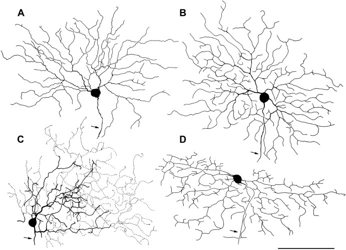
Figures


Similar articles
-
Retinal synaptic pathways underlying the response of the rabbit local edge detector.J Neurophysiol. 2010 May;103(5):2757-69. doi: 10.1152/jn.00987.2009. Epub 2010 Mar 24. J Neurophysiol. 2010. PMID: 20457864 Free PMC article.
-
GABA transporters regulate inhibition in the retina by limiting GABA(C) receptor activation.J Neurosci. 2002 Apr 15;22(8):3285-92. doi: 10.1523/JNEUROSCI.22-08-03285.2002. J Neurosci. 2002. PMID: 11943830 Free PMC article.
-
A novel GABA receptor modulates synaptic transmission from bipolar to ganglion and amacrine cells in the tiger salamander retina.J Neurosci. 1994 Mar;14(3 Pt 1):1213-23. doi: 10.1523/JNEUROSCI.14-03-01213.1994. J Neurosci. 1994. PMID: 7907138 Free PMC article.
-
GABAergic neurotransmission and retinal ganglion cell function.J Comp Physiol A Neuroethol Sens Neural Behav Physiol. 2015 Mar;201(3):261-83. doi: 10.1007/s00359-015-0981-z. Epub 2015 Feb 6. J Comp Physiol A Neuroethol Sens Neural Behav Physiol. 2015. PMID: 25656810 Review.
-
Receptor targets of amacrine cells.Vis Neurosci. 2012 Jan;29(1):11-29. doi: 10.1017/S0952523812000028. Vis Neurosci. 2012. PMID: 22310370 Free PMC article. Review.
Cited by
-
Role of Oxidative Stress in Ocular Diseases: A Balancing Act.Metabolites. 2023 Jan 27;13(2):187. doi: 10.3390/metabo13020187. Metabolites. 2023. PMID: 36837806 Free PMC article. Review.
-
Benzodiazepine and kainate receptor binding sites in the RCS rat retina.Graefes Arch Clin Exp Ophthalmol. 2003 Feb;241(2):154-60. doi: 10.1007/s00417-002-0611-7. Epub 2003 Jan 21. Graefes Arch Clin Exp Ophthalmol. 2003. PMID: 12605271
-
Two-photon imaging of nonlinear glutamate release dynamics at bipolar cell synapses in the mouse retina.J Neurosci. 2013 Jul 3;33(27):10972-85. doi: 10.1523/JNEUROSCI.1241-13.2013. J Neurosci. 2013. PMID: 23825403 Free PMC article.
-
Voltage-gated sodium channels improve contrast sensitivity of a retinal ganglion cell.J Neurosci. 2005 Aug 31;25(35):8097-103. doi: 10.1523/JNEUROSCI.1962-05.2005. J Neurosci. 2005. PMID: 16135767 Free PMC article.
-
The Retina: A Window into the Brain.Cells. 2021 Nov 23;10(12):3269. doi: 10.3390/cells10123269. Cells. 2021. PMID: 34943777 Free PMC article.
References
-
- Ammermüller J, Weiler R. Physiological and morphological characterization of OFF-center amacrine cells in the turtle retina. J Comp Neurol. 1988;273:137–148. - PubMed
-
- Amthor FR, Oyster CW, Takahashi EH. Morphology of ON-OFF direction-selective ganglion cells in the rabbit retina. Brain Res. 1984;298:187–190. - PubMed
-
- Amthor FR, Takahashi ES, Oyster CW. Morphologies of rabbit retinal ganglion cells with concentric receptive fields. J Comp Neurol. 1989a;280:72–96. - PubMed
-
- Amthor FR, Takahashi EH, Oyster CW. Morphologies of rabbit retinal ganglion cells with complex receptive fields. J Comp Neurol. 1989b;280:97–121. - PubMed
MeSH terms
Substances
LinkOut - more resources
Full Text Sources