. 2001 Jul 1;21(13):4915-22.
doi: 10.1523/JNEUROSCI.21-13-04915.2001.

Glutamatergic afferents from the hippocampus to the nucleus accumbens regulate activity of ventral tegmental area dopamine neurons

Affiliations

Glutamatergic afferents from the hippocampus to the nucleus accumbens regulate activity of ventral tegmental area dopamine neurons

S B Floresco et al. J Neurosci. .

Abstract

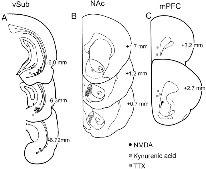
Several studies have shown that the mesolimbic dopamine (DA) system is strongly influenced by the ventral subiculum (vSub) of the hippocampus. To examine whether this occurs by activation of DA neuron firing, the effects of chemical stimulation of the vSub on ventral tegmental area (VTA) DA neuron activity were examined using extracellular single-unit recordings. Infusions of NMDA into the vSub increased the number of spontaneously firing DA cells recorded per electrode track, while having no effect on firing rate or burst firing. This response was abolished by intranucleus accumbens (NAc) infusions of the glutamate receptor antagonist kynurenic acid. This effect did not involve the prefrontal cortex, because infusions of tetrodotoxin into the prefrontal cortex did not affect the increase in spontaneously active DA cells. Infusions of either kynurenic acid into the NAc or tetrodotoxin into the vSub decreased the firing rate and burst firing of DA neurons without altering the number of spontaneously active DA neurons. These data show that glutamatergic afferents from the vSub to the NAc exert a potent excitatory effect on VTA DA neurons, influencing both DA neuron population activity and the regulation of the firing properties of these neurons. As a result, dysfunctions in hippocampal circuitries may contribute to the hyperexcitable state of the DA system that is present in schizophrenia.


Figures

Fig. 1.
Fig. 1.
Histological verification of all recording and infusion sites in the present study. The location of infusions is shown for all rats receiving the following: A, infusions of 0.75 μg of NMDA (black circles) or TTX (gray squares) into the vSub; B, infusions of kynurenic acid into the NAc (gray circles); or C, infusions of TTX into the mPFC (gray squares). Plates are taken from Paxinos and Watson (1997), and the numbers beside each plate correspond to millimeters from bregma.
Fig. 2.
Fig. 2.
Infusions of NMDA into the vSub increase DA neuron population activity. A, Activation of the vSub led to a dose-dependent increase in spontaneously active DA neurons. The mean number of spontaneously active DA neurons (+SEM) recorded per electrode track from rats receiving vehicle infusions (n = 6 rats; white bar), 0.25 or 0.75 μg of NMDA (n = 6 rats, 7 rats, respectively;black bars), or TTX (n = 6 rats;gray bar) into the vSub is shown. B, Blockade of glutamate transmission in the NAc prevented the activation of VTA DA neuron firing by vSub stimulation in a manner that was independent of the vSub–mPFC pathway. The mean number of spontaneously active DA neurons (+SEM) recorded per electrode track from rats receiving vehicle infusions (same as A; white bar), infusions of kynurenic acid (Kyn) into the NAc in combination with vehicle infusions into the vSub (n = 6 rats; hatched bar), infusions of kynurenic acid into the NAc in combination with 0.75 μg of NMDA into the vSub (n = 6 rats; cross-hatched bar), or infusions of TTX into the mPFC in combination with 0.75 μg of NMDA into the vSub (n = 5 rats;gray bar) is shown. *p < 0.05 and **p < 0.01 versus control; †p< 0.05 versus the group receiving 0.75 μg of NMDA.
Fig. 3.
Fig. 3.
Modulation of DA neuron firing rate by the vSub, the NAc, and the mPFC. A, In contrast to the effects on spontaneous activity, activation of the vSub did not change significantly the VTA DA neuron firing rate. Nonetheless, inactivation of the vSub did decrease VTA DA cell firing. The mean firing rate of DA neurons (+SEM) recorded from rats receiving vehicle infusions (white bar), 0.25 or 0.75 μg of NMDA (black bars), or TTX (gray bar) into the vSub is shown. B, Blockade of glutamate transmission in the NAc attenuates the DA neuron firing rate. The mean firing rate of DA neurons (+SEM) recorded from rats receiving vehicle infusions (same asA; white bar), infusions of kynurenic acid into the NAc in combination with vehicle infusions into the vSub (hatched bar), infusions of kynurenic acid into the NAc in combination with 0.75 μg of NMDA into the vSub (cross-hatched bar), or infusions of TTX into the mPFC in combination with 0.75 μg of NMDA into the vSub (gray bar) is shown. *p < 0.05 and **p < 0.01 versus control. C, Distribution of mean firing rates of all DA neurons recorded from rats receiving vehicle injection (C1), 0.25 μg of NMDA (C2) or 0.75 μg of NMDA (C3) into the vSub, or infusions of TTX into the mPFC in combination with 0.75 μg of NMDA into the vSub (C4) is shown. This suggests that the increase in DA cell spontaneous activity after vSub stimulation may be caused by the activation of slow-firing DA neurons.
Fig. 4.
Fig. 4.
Modulation of DA neuron burst firing by the vSub, the NAc, and the mPFC. A, As with firing rate, activation of the vSub did not cause a significant change in DA cell burst firing, although inactivation of the vSub did attenuate bursting. The mean % SIB of DA neurons (+SEM) recorded from rats receiving vehicle infusions (white bar), 0.25 or 0.75 μg of NMDA (black bars), or TTX (gray bar) into the vSub is shown. B, Similarly, glutamate transmission in the NAc also controls basal levels of burst firing of VTA DA neurons. The mean % SIB of DA neurons (+SEM) recorded from rats receiving vehicle infusions (same as A;white bar), infusions of kynurenic acid into the NAc in combination with vehicle infusions into the vSub (hatched bar), infusions of kynurenic acid into the NAc in combination with 0.75 μg of NMDA into the vSub (cross-hatched bar), or infusions of TTX into the mPFC in combination with 0.75 μg of NMDA into the vSub (gray bar) is shown. *p < 0.05 and **p < 0.01, versus control.C, Distribution of % SIB of all DA neurons recorded from rats receiving vehicle injection (C1) or infusions of TTX into the mPFC in combination with 0.75 μg of NMDA into the vSub (C2) is shown.
Fig. 5.
Fig. 5.
A model describing the neural circuitries by which the vSub may modulate DA neuron activity. Stimulation of the vSub by infusions of NMDA activates glutamatergic afferents to the NAc. This in turn is proposed to inhibit the activity of neurons in the VP. As a result, there is a removal of tonic inhibition of neurons in the VTA. In this model, we propose that the VP–VTA projection provides sufficient inhibitory influence to hold a subpopulation of DA neurons in an inactive state. Thus, inhibition of VP firing would lead to an increase in the number of VTA DA neurons firing spontaneously. The question mark beside the VP to VTA path indicates that, although this pathway is known to be present in the rat, its involvement in this response remains speculative at the present time.

References

    1. Bardgett ME, Henry JD. Locomotor activity and accumbens fos expression driven by ventral hippocampal stimulation require D1 and D2 receptors. Neuroscience. 1999;94:59–70. - PubMed
    1. Blaha CD, Allen LF, Das S, Ingles WL, Latimet MP, Vincent SR, Winn P. Modulation of dopamine efflux in the nucleus accumbens after cholinergic stimulation of the ventral tegmental area in intact, pedunculopontine tegmental nucleus-lesioned and laterodorsal tegmental nucleus-lesioned rats. J Neurosci. 1996;16:714–722. - PMC - PubMed
    1. Blaha CD, Yang CR, Floresco SB, Barr AM, Phillips AG. Stimulation of the ventral subiculum of the hippocampus evokes glutamate receptor-mediated changes in dopamine efflux in the rat nucleus accumbens. Eur J Neurosci. 1997;5:902–911. - PubMed
    1. Bogerts B. Recent advances in the neuropathology of schizophrenia. Schizophr Bull. 1993;19:431–445. - PubMed
    1. Brog JS, Salyapongse A, Deutch AY, Zahm DS. The patterns of afferent innervation of the core and shell in the “accumbens” part of the rat ventral striatum: immunohistochemical detection of retrogradely transported fluoro-gold. J Comp Neurol. 1993;338:255–278. - PubMed

Publication types

MeSH terms