

Behavioral functions of the reticular formation
- PMID: 114277
- PMCID: PMC8788642
- DOI: 10.1016/0165-0173(79)90017-1
Behavioral functions of the reticular formation
Abstract
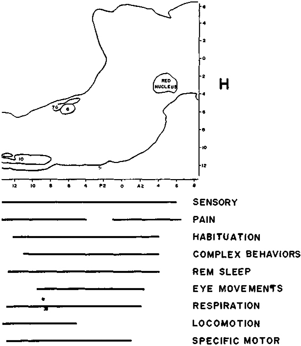
Studies of the behavioral correlates of activity in reticular formation cells, usually performed in restrained animals, have found units whose discharge relates to sensory stimuli, pain and escape behavior, conditioning and habituation, arousal, complex motivational states, REM sleep, eye movements, respiration and locomotion. Units with these different behavioral correlates were found in the same anatomical areas. Most studies report that a large proportion of encountered cells related to the behavior being studied. If one adds up the reported percentages, the total far exceeds 100%. Therefore it appears that many investigators are looking at the same cells and reaching very different conclusions about their behavioral roles. On the basis of observations in unrestrained cats, it is hypothesized that discharge in most RF cells is primarily related to the excitation of small groups of muscles. This hypothesis can parsimoniously explain many previous observations on the behavioral correlates of these cells, and is consistent with anatomical, physiological and phylogenetic studies of the reticular formation. The hypothesized simplicity of reticular formation unit function is contrasted with the complexity of the behavioral functions mediated by the RF, and the implications of this contrast discussed.
Figures
Similar articles
-
Pontine reticular formation neurons: relationship of discharge to motor activity.Science. 1977 May 6;196(4290):678-80. doi: 10.1126/science.193185. Science. 1977. PMID: 193185 Free PMC article.
-
Interrelationship among multiunit activity of the midbrain reticular formation and lateral geniculate nucleus, thalamocortical arousal, and behavior in rats.J Comp Physiol Psychol. 1975 Apr;89(2):131-57. doi: 10.1037/h0076651. J Comp Physiol Psychol. 1975. PMID: 1133235
-
Mechanisms of habituation in the brain stem.Psychol Rev. 1972 May;79(3):237-44. doi: 10.1037/h0032689. Psychol Rev. 1972. PMID: 4560358 Review. No abstract available.
-
Habituation: electrical changes in the visual system.Neuropsychologia. 1972 Apr;10(1):125-32. doi: 10.1016/0028-3932(72)90050-4. Neuropsychologia. 1972. PMID: 4624748 No abstract available.
-
[Control mechanisms of locomotor movements from a viewpoint of behavioral control].Nihon Shinkei Seishin Yakurigaku Zasshi. 1995 Jun;15(3):281-7. Nihon Shinkei Seishin Yakurigaku Zasshi. 1995. PMID: 7584722 Review. Japanese.
Cited by
-
Diversity of reticulospinal systems in mammals.Curr Opin Physiol. 2019 Apr;8:161-169. doi: 10.1016/j.cophys.2019.03.001. Epub 2019 Mar 12. Curr Opin Physiol. 2019. PMID: 31763514 Free PMC article.
-
Phylogeny and the function of REM sleep.Behav Brain Res. 1995 Jul-Aug;69(1-2):29-34. doi: 10.1016/0166-4328(95)00023-m. Behav Brain Res. 1995. PMID: 7546315 Free PMC article. Review.
-
Behavioral organization of reticular formation: studies in the unrestrained cat. I. Cells related to axial, limb, eye, and other movements.J Neurophysiol. 1983 Sep;50(3):696-716. doi: 10.1152/jn.1983.50.3.696. J Neurophysiol. 1983. PMID: 6619914 Free PMC article.
-
Electrophysiological study of supraspinal input and spinal output of cat's subnucleus reticularis dorsalis (SRD) neurons.PLoS One. 2013;8(3):e60686. doi: 10.1371/journal.pone.0060686. Epub 2013 Mar 27. PLoS One. 2013. PMID: 23544161 Free PMC article.
-
Human hypocretin and melanin-concentrating hormone levels are linked to emotion and social interaction.Nat Commun. 2013;4:1547. doi: 10.1038/ncomms2461. Nat Commun. 2013. PMID: 23462990 Free PMC article.
References
-
- Adams DB, The activity of single cells in the midbrain and hypothalamus of the cat during affective defense behavior, Arch. ital. Biol, 106 (1968) 243–269. - PubMed
-
- Adams DB, Cells related to fighting behavior recorded from mid-brain central grey neuropil of cat, Science, 159 (1968) 894–896. - PubMed
-
- Amassian VE and Devito RV, Unit activity in reticular formation and nearby structures, J. Neurophysiol, 17 (1954) 575–603. - PubMed
-
- Anderson ME, Yoshida M and Wilson VS, Tectal and tegmental influences on cat forelimb and hindlimb motorneurons, J. Neurophysiol, 35 (1972) 462–470. - PubMed
-
- Appleberg B and Jenskog T, Mesencephalic fusimotor control, Exp. Brain Res, 15 (1972) 97–112. - PubMed
Publication types
MeSH terms
Grants and funding
LinkOut - more resources
Full Text Sources
Miscellaneous