. 2001 Jul 3;98(14):8071-6.
doi: 10.1073/pnas.141215898. Epub 2001 Jun 26.

Different modes of hippocampal plasticity in response to estrogen in young and aged female rats

Affiliations

Different modes of hippocampal plasticity in response to estrogen in young and aged female rats

M M Adams et al. Proc Natl Acad Sci U S A. .

Abstract

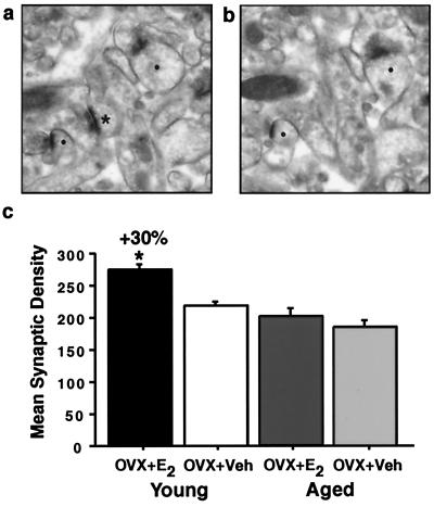
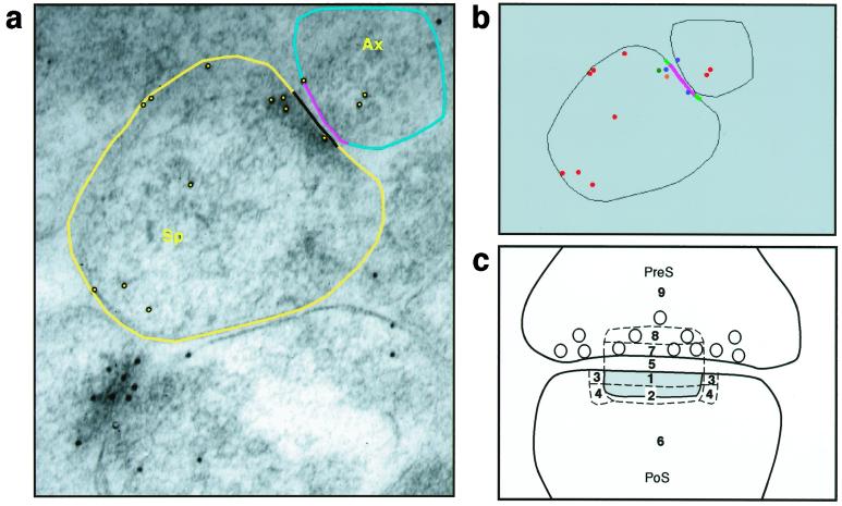
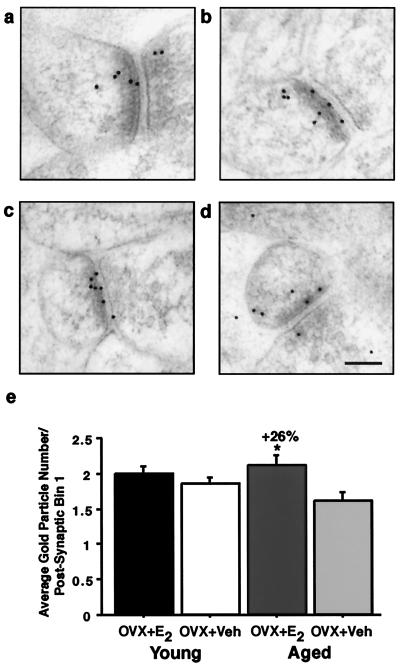
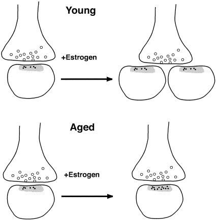
Estrogen regulates hippocampal dendritic spine density and synapse number in an N-methyl-D-aspartate (NMDA) receptor-dependent manner, and these effects may be of particular importance in the context of age-related changes in endocrine status. We investigated estrogen's effects on axospinous synapse density and the synaptic distribution of the NMDA receptor subunit, NR1, within the context of aging. Although estrogen induced an increase in axospinous synapse density in young animals, it did not alter the synaptic representation of NR1, in that the amount of NR1 per synapse was equivalent across groups. Estrogen replacement in aged female rats failed to increase axospinous synapse density; however, estrogen up-regulated synaptic NR1 compared with aged animals with no estrogen. Therefore, the young and aged hippocampi react differently to estrogen replacement, with the aged animals unable to mount a plasticity response generating additional synapses, yet responsive to estrogen with respect to additional NMDA receptor content per synapse. These findings have important implications for estrogen replacement therapy in the context of aging.


Figures

Figure 1
Figure 1
Synapse density in stratum radiatum of CA1 in young and aged estrogen (OVX + E2)- and vehicle (OVX + Veh)-treated groups. Ultrastructural images from a representative young animal illustrating the disector method. For each animal, 15 pairs of adjacent digital images were taken. One section was considered the reference (a) and the other the look-up (b). Only synaptic profiles (*) contained in the reference but not in the corresponding look-up planes were counted. [Note: (●), Solid circles denote profiles contained in both planes.] This procedure was repeated by switching the reference and look-up sections. (c) Analysis revealed a significant increase in young OVX + E2 compared with OVX + Veh (P < 0.005) but no difference between aged OVX + E2 and OVX + Veh (P > 0.38). The young OVX + E2 were significantly different from both aged groups (both P values < 0.0001).
Figure 2
Figure 2
Bin analysis of synaptic NR1. Images illustrate software used for this analysis. (a) Outlines of the relevant synaptic components, i.e., presynaptic and postsynaptic membranes, length of the postsynaptic density (PSD), and length of the presynaptic membrane that corresponds to the length of the PSD. (b) The program provides a data map of the distribution of gold particles relative to these membranes. (c) A schematic diagram of different postsynaptic (PoS) compartments. Two major postsynaptic bins were designated: Bin 1 is 0–30 nm from the inner leaflet of the postsynaptic membrane, and Bin 2 is 30–60 nm from the postsynaptic membrane. Bins 3 and 4 are side bins, 15 nm lateral to Bins 1 and 2. Bin 5 is the synaptic cleft; Bin 6 is a cytoplasmic bin that includes gold particles >60 nm from the postsynaptic membrane. Two major presynaptic (PreS) zones were designated: Bin 7 is 0–30 nm from the inner border of the presynaptic membrane; Bin 8 is 30–60 nm from the presynaptic membrane. Bin 9 is a presynaptic bin that includes gold >60 nm from the presynaptic membrane. Sp, spine; Ax, Axon.
Figure 3
Figure 3
Bin analysis of NR1 in young and aged OVX + E2 and OVX + Veh. Images from young (a and b) and aged (c and d) animals illustrating the distribution of the NR1 postembedding immunogold. (e) Analysis revealed no significant differences in the synaptic distribution of NR1 between the young (a) OVX + E2 and (b) OVX + Veh (P > 0.15). However, the aged (c) OVX + E2 had significantly more NR1 per postsynaptic Bin 1 compared with (d) OVX + Veh (P < 0.05). (Bar = 100 nm.)
Figure 4
Figure 4
A schematic of estrogen-induced plasticity in young and aged animals. Estrogen treatment increases NR1 expression per synapse in aged hippocampus, whereas it increases spine number but not synaptic NR1 in young female rat hippocampus. Small black dots represent immunogold particles labeling NR1 associated with postsynaptic Bin 1; open circles are synaptic vesicles; the gray zone indicates the postsynaptic density.

References

    1. Lamberts S W J, van den Beld A W, van der Lely A-J. Science. 1997;278:419–424. - PubMed
    1. Fink G. Sci Prog. 1986;70:403–423. - PubMed
    1. Woolley C S. Horm Behav. 1998;34:140–148. - PubMed
    1. Sherwin B B. Neurology. 1997;48:S21–S26. - PubMed
    1. Berry B, McMahan R, Gallagher M. Behav Neurosci. 1997;111:267–274. - PubMed

Publication types