

Identification of a subpopulation of rapidly self-renewing and multipotential adult stem cells in colonies of human marrow stromal cells
- PMID: 11427725
- PMCID: PMC35429
- DOI: 10.1073/pnas.141221698
Identification of a subpopulation of rapidly self-renewing and multipotential adult stem cells in colonies of human marrow stromal cells
Abstract
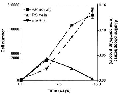
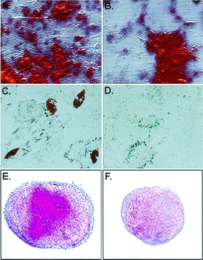
Marrow stromal cells are adult stem cells from bone marrow that can differentiate into multiple nonhematopoietic cell lineages. Previous reports demonstrated that single-cell-derived colonies of marrow stromal cells contained two morphologically distinct cell types: spindle-shaped cells and large flat cells. Here we found that early colonies also contain a third kind of cell: very small round cells that rapidly self-renew. Samples enriched for the small cells had a greater potential for multipotential differentiation than samples enriched for the large cells. Also, the small cells expressed a series of surface epitopes and other proteins that potentially can be used to distinguish the small cells from the large cells. The results suggested it will be important to distinguish the major subpopulations of marrow stromal cells in defining their biology and their potential for cell and gene therapy.
Figures
References
-
- Friedenstein A J, Chailakhjan R K, Lalykina K S. Cell Tissue Kinet. 1970;3:393–403. - PubMed
-
- Castro-Malaspina H, Gay R E, Resnick G, Kapoor N, Meyers P, Chiarieri D, McKenzie S, Broxmeyer H E, Moore M A. Blood. 1980;56:289–301. - PubMed
-
- Mets T, Verdonk G. Mech Ageing Dev. 1981;16:81–89. - PubMed
-
- Piersma A H, Brockbank K G, Ploemacher R E, van Vliet E, Brakel-van Peer K M, Visser P J. Exp Hematol. 1985;13:237–243. - PubMed
-
- Owen M E, Friedenstein A J. Cell and Molecular Biology of Vertebrate Hard Tissues. Chichester, UK: Ciba Foundation Symposium; 1988. pp. 42–60. - PubMed
Publication types
MeSH terms
Grants and funding
LinkOut - more resources
Full Text Sources
Other Literature Sources
Medical
