. 2001 Jul 3;98(14):7783-8.
doi: 10.1073/pnas.141224398. Epub 2001 Jun 26.

Raf-1 promotes cell survival by antagonizing apoptosis signal-regulating kinase 1 through a MEK-ERK independent mechanism

Affiliations

Raf-1 promotes cell survival by antagonizing apoptosis signal-regulating kinase 1 through a MEK-ERK independent mechanism

J Chen et al. Proc Natl Acad Sci U S A. .

Abstract

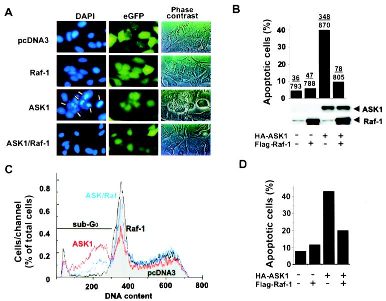
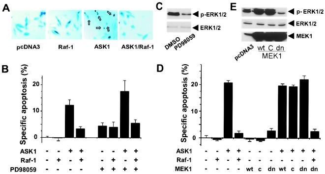
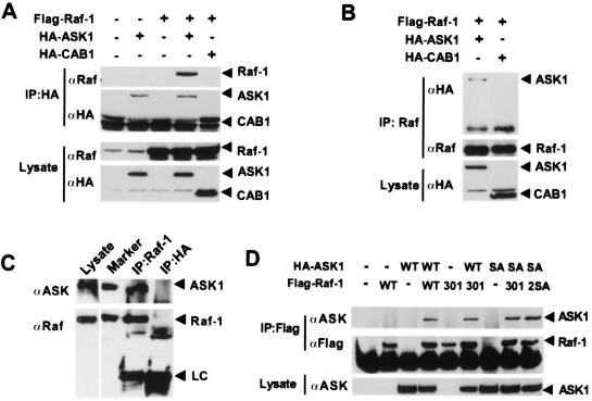
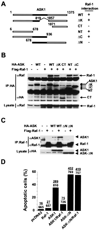
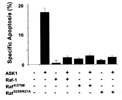
The Ser/Thr kinase Raf-1 is a protooncogene product that is a central component in many signaling pathways involved in normal cell growth and oncogenic transformation. Upon activation, Raf-1 phosphorylates mitogen-activated protein kinase kinase (MEK), which in turn activates mitogen-activated protein kinase/extracellular signal-regulated kinases (MAPK/ERKs), leading to the propagation of signals. Depending on specific stimuli and cellular environment, the Raf-1--MEK--ERK cascade regulates diverse cellular processes such as proliferation, differentiation, and apoptosis. Here, we describe a MEK--ERK-independent prosurvival function of Raf-1. We found that Raf-1 interacts with the proapoptotic, stress-activated protein kinase ASK1 (apoptosis signal-regulating kinase 1) in vitro and in vivo. Deletion analysis localized the Raf-1 binding site to the N-terminal regulatory fragment of ASK1. This interaction allows Raf-1 to act independently of the MEK--ERK pathway to inhibit apoptosis. Furthermore, catalytically inactive forms of Raf-1 can mimic the wild-type effect, raising the possibility of a kinase-independent function of Raf-1. Thus, Raf-1 may promote cell survival through its protein-protein interactions in addition to its established MEK kinase function.


Figures

Figure 1
Figure 1
Expression of Raf-1 inhibits ASK1-induced apoptosis. HeLa cells were transfected with the plasmids pcDNA3–HA–ASK1 (1.2 μg) or pcDNA3–FLAG–Raf-1 (0.4 μg) along with an eGFP expression vector (0.4 μg) as indicated. Eighteen hours posttransfection, cells were placed in serum-free medium for an additional 24 h before staining with DAPI. Nuclear morphology of transfected cells was examined by fluorescence microscopy as described (18), and apoptotic nuclei are indicated by arrows (A). The fraction of transfected cells with fragmented nuclei was quantified in a blind manner (B Upper). Cell lysates from each sample were subjected to SDS/PAGE and Western blotting with anti-Raf-1 (SC133; Santa Cruz Biotechnology) or anti-HA (12CA5) antibodies (B Lower). (C) COS7 cells were transfected with plasmids as in A along with an eGFP-F expression vector. Twenty-four hours after transfection, total cells were harvested, their DNA was stained with propidium iodide, and eGFP and propidium iodide signals were measured on a FACSort flow cytometer. Transfected cells (eGFP-positive) were placed in various phases of the cell cycle based on their DNA content. Apoptotic cells with fragmented DNA (subG0) are indicated. (D) Data from C were compiled to show the amount of apoptosis caused by expression of transfected plasmids.
Figure 2
Figure 2
MEK activity is not required for Raf-1 to inhibit ASK1. (A) Morphology of ASK1 transfected cells. HeLa cells were cotransfected with a β-gal expression vector and test plasmids as indicated. Twenty-four hours posttransfection, cells were fixed and stained with 5-bromo-4-chloro-3-indolyl-β-D-galactopyranoside. Shrunken apoptotic cells with rounded-up shape were scored as apoptotic (arrows). (B) Effect of MEK inhibition by PD98059 on Raf-1 function. Six hours after transfection as in A, HeLa cells were treated with PD98059 (60 μM) or vehicle (DMSO) for 18 h before being stained with 5-bromo-4-chloro-3-indolyl-β-D-galactopyranoside and scored for apoptosis in a blind fashion. Specific apoptosis is derived by subtracting the level of apoptosis seen in pcDNA3-transfected cells. At least 500 cells were scored for each sample. Results shown are representative of three independent experiments. (C) Inhibition of ERK1/2 activation by PD98059. Cell lysates from HeLa cells treated with 60 μM PD98059 or vehicle were probed by Western blotting with either an ERK1/2 activation-specific antibody (Upper; Cell Signaling Technology) or pan-ERK1/2 antibody (Lower; Cell Signaling Technology). (D) Effect of MEK1 overexpression on ASK1-induced apoptosis. HeLa cells were transfected with a β-gal reporter together with indicated expression vectors for 12 h, serum-starved for 24 h, and scored for apoptosis as in A. Data are summary of three independent experiments. (E) Effect of MEK1 expression on ERK1/2 activation. Lysates from HeLa cells treated as in D were subjected to SDS/PAGE and Western blotting with ERK1/2 activation-specific or pan antibodies or anti-MEK antibody (Santa Cruz Biotechnology).
Figure 3
Figure 3
Raf-1 specifically interacts with ASK1. (A) HA–ASK1 immunocomplex contains Raf-1. COS7 cells were transfected with the expression vector for HA–ASK1 or HA–CAB1 together with FLAG–Raf-1 (18). After 48 h, cell lysates were prepared, and ASK1 or CAB1 was immunoprecipitated with anti-HA antibody. Immunoprecipitates were washed extensively with Nonidet P-40 (1%) lysis buffer before Western blotting with antibodies to Raf-1 and HA (Upper). (Lower) Expression levels of Raf-1 and HA-ASK1 or HA-CAB1 in the lysates. (B) Raf-1 immunocomplexes contain ASK1. Polyclonal anti-Raf-1 antibody (Santa Cruz Biotechnology) was used to immunoprecipitate Raf-1 as in A. HA-ASK1 was detected in the Raf-1 immunoprecipitates with anti-HA antibody. (C) Endogenous Raf-1 and ASK1 form complexes in L929 cells. Immunoprecipitates were prepared from L929 cell lysates (left lane) using either anti-Raf-1 monoclonal antibody (Transduction Laboratories) or anti-HA antibody as a negative control and probed for ASK1 using the antibody DAV (ref. ; Upper). Coimmunoprecipitated ASK1 and Raf-1 comigrate, respectively, with overexpressed HA–ASK1 and FLAG–Raf-1 (Marker lane). Antibody light chains (LC) in the immunoprecipitates are indicated. (D) 14-3-3 binding and Raf-1 kinase activity are not required for the Raf-1–ASK1 interaction. COS7 cells were cotransfected with plasmids encoding FLAG–Raf-1WT, catalytically inactive FLAG–RafK375M (301), or 14-3-3 binding defective FLAG–RafS259/621A (2SA) and HA-ASK1WT or 14-3-3 binding defective HA–ASK1S967A (SA). FLAG–Raf-1 complex was precipitated by using anti-FLAG antibody (Sigma) and probed with anti-ASK1 antibody (Santa Cruz Biotechnology). Expression levels of HA–ASK1 were verified by Western blotting (Lower). As a control, FLAG–RafS259/621A was found to bind HA–ASK1WT (data not shown).
Figure 4
Figure 4
The N-terminal domain of ASK1 mediates the Raf-1 interaction. (A) Schematic diagram of ASK1 proteins. The shaded portion of the boxes represents the ASK1 kinase domain. Association of ASK1 mutants with Raf-1 is summarized. (B) The N-terminal domain of ASK1 is required for Raf-1 binding. FLAG–Raf-1 was transiently transfected into COS7 cells with HA–ASK1WT or truncated mutants. HA–ASK1 protein complexes were immunoprecipitated and subjected to SDS/PAGE and Western blotting with anti-HA (Middle) and anti-Raf-1 antibodies (Top). Lysates from each sample were probed with anti-Raf-1 antibodies (Bottom). (C) Raf-1 does not interact with ASK1-ΔN. Raf-1 protein complexes were immunoprecipitated from each sample with anti-Raf-1 antibody and subjected to SDS/PAGE and Western blotting with anti-ASK1 antibody. Overexposure shows the interaction of endogenous Raf-1 with overexpressed HA–ASK1, but even overexpressed Raf-1 was incapable of binding to ASK1-ΔN. (D) Raf-1 cannot block ASK1-ΔN induced apoptosis. HeLa cells were transfected with plasmids as indicated together with an eGFP marker vector. The nuclear morphology-based assay described in Fig. 1 was used to score for apoptotic cells.
Figure 5
Figure 5
Raf-1 inhibits ASK1-induced apoptosis independently of its catalytic function. A HeLa cell morphology-based assay as in Fig. 2 was used to score for specific apoptosis. Plasmids used were the same as described in Fig. 3C.

References

    1. Morrison D K, Cutler R E. Curr Opin Cell Biol. 1997;9:174–179. - PubMed
    1. Kolch W. Biochem J. 2000;351:289–305. - PMC - PubMed
    1. Cleveland J L, Troppmair J, Packham G, Askew D S, Lloyd P, Gonzalez-Garcia M, Nunez G, Ihle J N, Rapp U R. Oncogene. 1994;9:2217–2226. - PubMed
    1. Xia Z, Dickens M, Raingeaud J, Davis R J, Greenberg M E. Science. 1995;270:1326–1331. - PubMed
    1. Erhardt P, Schremser E J, Cooper G M. Mol Cell Biol. 1999;19:5308–5315. - PMC - PubMed

Publication types

Substances