. 2001 Jul 3;98(14):7970-5.
doi: 10.1073/pnas.141224998. Epub 2001 Jun 26.

Onset of natural killer cell lymphomas in transgenic mice carrying a truncated HMGI-C gene by the chronic stimulation of the IL-2 and IL-15 pathway

Affiliations

Onset of natural killer cell lymphomas in transgenic mice carrying a truncated HMGI-C gene by the chronic stimulation of the IL-2 and IL-15 pathway

G Baldassarre et al. Proc Natl Acad Sci U S A. .

Abstract

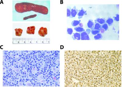
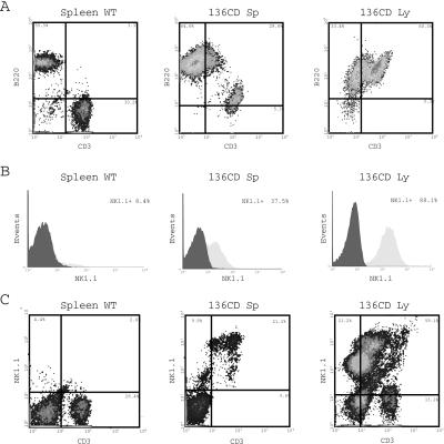
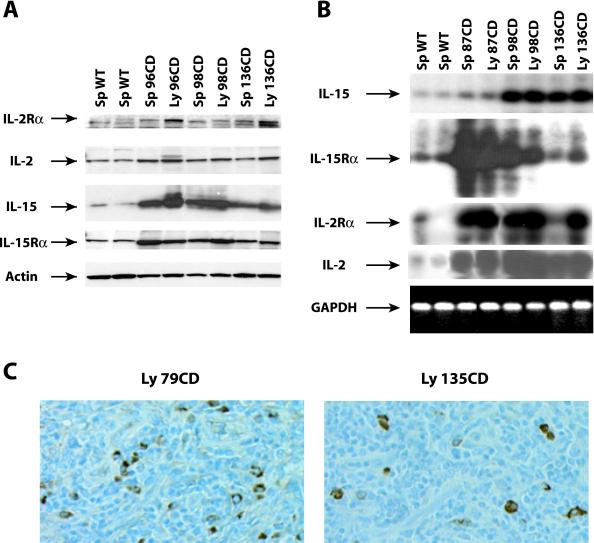
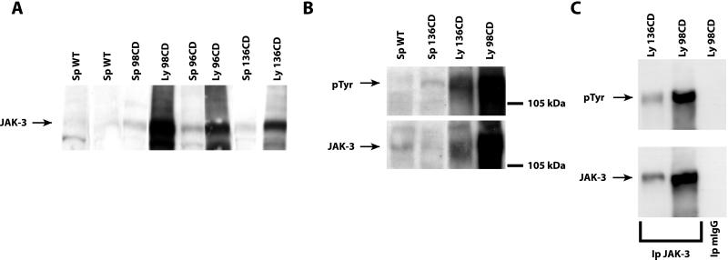
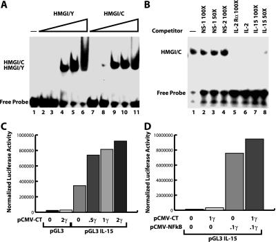
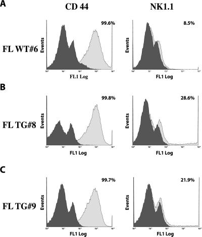
Rearrangements of the high mobility group protein I-C (HMGI-C) gene, consisting in the loss of the carboxyl-terminal tail, have been frequently detected in benign human tumors of mesenchymal origin. We have previously demonstrated that transgenic (TG) mice carrying a truncated HMGI-C construct (HMGI-C/T) exhibit a giant phenotype together with a predominantly abdominal/pelvic lipomatosis. Here, we report that HMGI-C/T TG mice develop natural killer (NK)-T/NK cell lymphomas starting from 12 months of age. We found an increased expression of IL-2 and IL-15 proteins and their receptors in these lymphomas, and we demonstrate that HMGI-C/T protein positively regulates their expression in vitro. Therefore, the HMGI-C/T-mediated chronic stimulation of the IL-2/IL-15 pathway could be responsible for the onset of NK-T/NK cell lymphomas in HMGI-C/T TG mice.


Figures

Figure 1
Figure 1
HMGI-C/T TG mice develop large cell T lymphomas. (A Upper) Comparison between normal and pathological spleens. (Lower) Enlargement of pathological lymph nodes in TG mice. (B) Cytospin analysis of pathological lymphocytes extracted from laterocervical lymph node. The large cell phenotype and the presence of azurophilic granules are showed. (C) Hematoxylin-eosin staining of a pathological lymph node revealed the presence of large cells. (D) CD3 stained positively in lymphoid cells. In each case, a representative field is shown.
Figure 2
Figure 2
HMGI-C/T TG mice develop T/NK cell lymphomas. (A) CD3 and B220 expression on lymphocytes extracted from a WT mouse (Left), a pathological spleen (Center), or a pathological lymph node (Right). The percentage of CD3+, B220+, and CD3+/B220+ is reported in the figure. (B) Expression of NK1.1 antigen in WT splenocytes (Left), TG splenocytes (Center), and pathological lymph node (Right). The percentage of NK1.1 positive cells is reported. (C) CD3 and NK1.1 expression in normal (Left) and pathological spleen (Center) and lymph node (Right).
Figure 3
Figure 3
Overexpression of IL-2 and IL-15 and their receptors in NK-T/NK cells derived from HMGI-C/T TG mice. (A) Expression of IL-2 Rα, IL-2, IL-15, and IL-15 Rα proteins in two different control spleens and in six different pathological specimens. The same Western blot was incubated with antibody vs. actin protein to normalize the amount of loaded proteins. (B) RT-PCR analysis of RNA extracted from two different WT spleens and six pathological specimens. The expression of IL-15, IL-15 Rα, IL-2, and IL-2 Rα is shown. The glyceraldehyde-3-phosphate dehydrogenase (GAPDH) expression was determined as an internal control of RNA quantity and status. (C) IL-15 expression analyzed by immunohistochemistry in two different pathological lymph nodes.
Figure 4
Figure 4
Levels and phosphorylation status of Jak-3 protein in WT and pathological specimens. (A) Western blot analysis determining the expression of Jak-3 protein in two different control spleens and in six different pathological specimens. (B) Phosphotyrosinse expression in one WT spleen and three different pathological specimens. The same Western blot was incubated with antibody vs. the Jak-3 protein. (C) Immunoprecipitation of Jak-3 protein in two different lymphomatous samples. The same blot was hybridized with anti-pTyr antibody and then stripped and reprobed with anti Jak-3 Ab. Normal mouse IgGs (mIgG) were used as a negative control.
Figure 5
Figure 5
HMGI-C/T protein binds to and activates the IL-15 promoter. (A) IL-15 −84 to −52 oligonucleotide recognized by increasing amounts (5, 10, 20, 50, and 100 ng, respectively) of HMGI/Y (lanes 2–6) or HMGI/C recombinant protein (lanes 8–11). (B) In vitro binding of HMGI-C protein to the IL-15 oligonucleotide competed with 30 ng of HMGI/C recombinant protein alone (lane 2) or in presence of two unrelated oligonucleotides (lanes 3 and 4). Specific competition of DNA protein interaction was obtained with a 50- to 100-fold excess of IL-2 Rα (lane 5), IL-2 (lane 6), or IL-15 (lanes 7 and 8) cold oligonucleotides. (C) Luciferase assay using the IL-15 promoter region transfected alone or with increasing concentrations of pCMV-HMGI-C/T vector. (D) Luciferase assay using the IL-15 promoter region transfected alone or with pCMV-NF-κB alone or in combination with pCMV-HMGI-C/T vector. In all cases, a typical experiment is reported.
Figure 6
Figure 6
Increasing expression of NK1.1 antigen on cultured fetal lymphocyte-derived HMGI-C/T TG embryos. CD44 (Left) and NK1.1 (Right) expression on fetal lymphocytes extracted from a WT embryo (A), and two different HMGI-C/T TG embryos (B and C). The percentage of CD44+ and NK1.1+ is reported in the figure.

References

    1. Manfioletti G, Giancotti V, Bandiera A, Buratti E, Sautiere P, Cary P, Crane-Robinson C, Coles B, Goodwin G H. Nucleic Acids Res. 1991;19:6793–6797. - PMC - PubMed
    1. Goodwin G, Bustin M. In: Architecture of Eukaryotic Genes. Kahl G, editor. Weinheim, Germany: VCH; 1988. pp. 187–205.
    1. Thanos D, Maniatis T. Cell. 1992;71:777–789. - PubMed
    1. Grosschedl R, Giese K, Pagel J. Trends Genet. 1994;10:94–100. - PubMed
    1. Mantovani F, Covaceuszach S, Rustighi A, Sgarra R, Heath C, Goodwin G H, Manfioletti G. Nucleic Acids Res. 1998;26:1433–1439. - PMC - PubMed

Publication types

MeSH terms

LinkOut - more resources