. 2001 Jul 3;98(14):7869-74.
doi: 10.1073/pnas.141226798. Epub 2001 Jun 26.

Xenopus Bcl-X(L) selectively protects Rohon-Beard neurons from metamorphic degeneration

Affiliations

Xenopus Bcl-X(L) selectively protects Rohon-Beard neurons from metamorphic degeneration

L Coen et al. Proc Natl Acad Sci U S A. .

Abstract

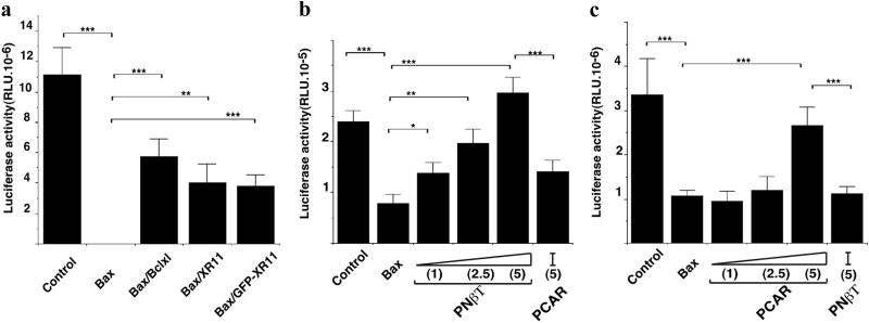
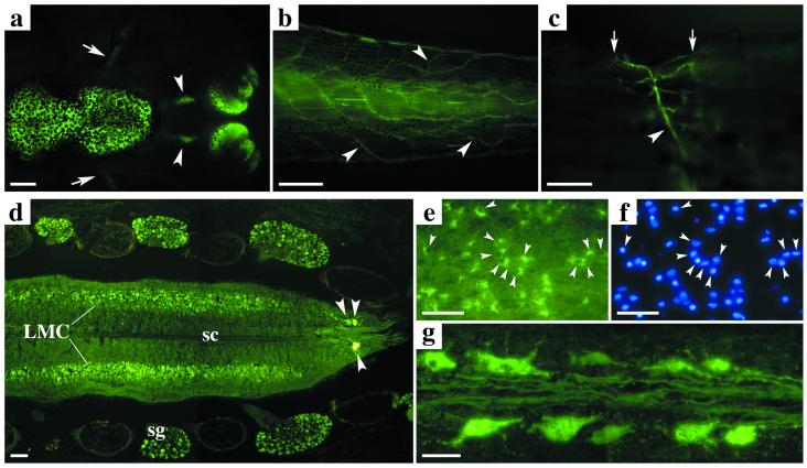
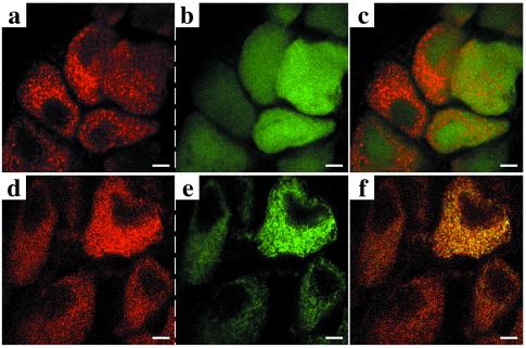
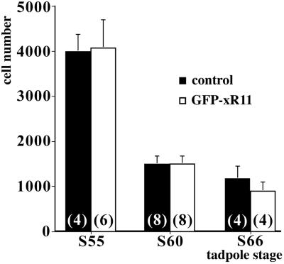
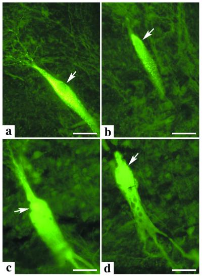
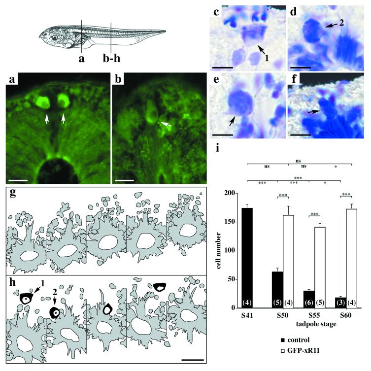
Amphibian metamorphosis involves extensive, but selective, neuronal death and turnover, thus sharing many features with mammalian postnatal development. The antiapoptotic protein Bcl-X(L) plays an important role in postnatal mammalian neuronal survival. It is therefore of interest that accumulation of the mRNA encoding the Xenopus Bcl-X(L) homologue, termed xR11, increases abruptly in the nervous system, but not in other tissues, during metamorphosis in Xenopus tadpoles. This observation raises the intriguing possibility that xR11 selectively regulates neuronal survival during postembryonic development. To investigate this hypothesis, we overexpressed xR11 in vivo as a green fluorescent protein (GFP)-xR11 fusion protein by using somatic and germinal transgenesis. Somatic gene transfer showed that the fusion protein was effective in counteracting, in a dose-dependent manner, the proapoptotic effects of coexpressed Bax. When GFP-xR11 was expressed from the neuronal beta-tubulin promoter by germinal transgenesis we observed neuronal specific expression that was maintained throughout metamorphosis and beyond, into juvenile and adult stages. Confocal microscopy showed GFP-xR11 to be exclusively localized in the mitochondria. Our findings show that GFP-xR11 significantly prolonged Rohon-Beard neuron survival up to the climax of metamorphosis, even in the regressing tadpole tail, whereas in controls these neurons disappeared in early metamorphosis. However, GFP-xR11 expression did not modify the fate of spinal cord motoneurons. The selective protection of Rohon-Beard neurons reveals cell-specific apoptotic pathways and offers approaches to further analyze programmed neuronal turnover during postembryonic development.


Figures

Figure 1
Figure 1
Validation of the antiapoptotic effect of xR11 and GFP-xR11 and the promoter specificity of constructs by somatic gene transfer. (a) Cell survival was measured by the luciferase activity (RLU) in muscle extracts 48 h after transfection of 1 μg pcDNA3-LUC into the dorsal muscle of stage 56 tadpoles. The following plasmids were coinjected with pcDNA3-LUC (from left to right): 2 μg pcDNA3 (empty vector, control), 1 μg pcDNA3-Bax, 1 μg pcDNA3-Bax with 1 μg CMV-Bcl-XL, 1 μg pcDNA3-Bax with 1 μg pcDNA3-xR11, 1 μg pcDNA3-Bax with 1 μg peGFP-xR11. All cDNA were CMV-driven. The total amount of DNA injected was brought up to 3 μg by adding control pcDNA3 where necessary. Note that GFP-xR11 is as potent as xR11 and Bcl-XL in counteracting the apoptotic effects of human BAX. (b) Cell survival was measured with tissue-specific promoters in brain extracts 48 h after transfection. The total DNA used was made up to 450 ng, using control vector and complexed with polyethyleneimine. In addition to 150 ng pcDNA3-LUC (control, far left), the following plasmids were coinjected (from left to right): 50 ng pcDNA3-Bax, 50 ng pcDNA3-Bax with 50 ng t-GFP-xR11, 50 ng pcDNA3-Bax with 2.5× t-GFP-xR11, 50 ng pcDNA3-Bax with 5× excess t-GFP-xR11, 50 ng pcDNA3-Bax with 5× excess pCAR-GFP-xR11. Note that in the brain, Bax-dependent apoptosis is only blocked when GFP-xR11 is expressed from the Nβt, but not the pCAR promoter. (c) Cell survival was measured with tissue-specific promoters in muscle extracts 48 h after transfection. Total DNA transfected (as free DNA) was brought up to 2.8 μg, using control vector where necessary. In addition to 1 μg pcDNA3-LUC (control, far left), the following plasmids were coinjected (from left to right): 300 ng pcDNA3-Bax, 300 ng pcDNA3-Bax with 300 ng pCAR-GFP-xR11, 300 ng pcDNA3-Bax with 2.5× excess pCAR-GFP-xR11, 300 ng of pcDNA3-Bax with 5× excess pCAR-GFP-xR11, 300 ng pcDNA3-Bax with 5× excess t-GFP-xR11. Note that in muscle, sufficient GFP-xR11 is expressed from the pCAR, but not the Nβt promoter, to block Bax-dependent apoptosis. Means ± SEM are given; n > 9 in each group. *, P < 0.05; **, P < 0.01; ***, P < 0.001.
Figure 2
Figure 2
GFP and GFP-xR11 expression from the Nβt promoter in transgenic Xenopus is limited to the nervous system. (a–c) In toto GFP expression from a Nβt-GFP stage 60 tadpole: (a) brain, olfactory nerves (arrowheads), olfactory bulbs, and optic nerves (arrows); (b) spinal cord and nerves (arrowheads) innervating the tail; (c) neuromuscular junctions (arrows) and axons (arrowhead). (d–g) Longitudinal cryostat sections (20 μm) of spinal cord from a Nβt-GFP-xR11 animal at climax (stage 61): (d) spinal ganglia, interneurons, primary motoneurons (arrowheads), and lateral columns of secondary motoneurons (LMC); (e and f) all interneurons express GFP (compare GFP in e with Hoechst labeling in f); (g) spinal cord primary motoneurons and their axons. LMC: lateral motor column; sg: spinal ganglion; sc: spinal cord. [Scale bars = 1 mm (a and b), 100 μm (c and d), and 50 μm (e-g).]
Figure 3
Figure 3
GFP-xR11, but not GFP alone, is localized in mitochondria. (a–f) Confocal analysis on transversal cryostat sections (30 μm) with a Cy3-coupled anti-cytochrome c (mitochondrial-specific marker) shows a punctuate pattern (a and d) colocalizing with GFP-xR11 (e), but not with GFP alone (b), which gives a homogenous, diffuse signal. Superposition of Cy3 and GFP signal (c) or GFP-xR11 signal (f) is shown in spinal ganglion neurons from a postmetamorphic (stage 66) animal. [Scale bars = 5 μm.]
Figure 4
Figure 4
GFP-xR11 expression does not abrogate motoneuron loss in the lumbar lateral motor column (L-LMC) during metamorphosis. Motoneurons were counted in the left and right L-LMC in animals expressing GFP or GFP-xR11 at stages (S) 55, 60, and 66. Means ± SD are shown (the n for each group is indicated at the base of the histogram). No differences between GFP or GFP-xR11 animals were seen at any stage (P > 0.05 for each comparison).
Figure 5
Figure 5
M neuron morphology and fate are modified by expression of GFP-xR11. M neurons from GFP-xR11 (a and b) or GFP (c and d) animals at stages 61 (a and c) and 66 (b and d). Transversal sections (30 μm) of hindbrain are shown with arrows indicating each M cell nucleus. At metamorphic climax (stage 61) the GFP-xR11 M (a) has a regular appearance whereas the GFP M (c) has a spreading cytoplasm with small vacuoles and an irregular membrane. On completion of metamorphosis (stage 66) these differences are more pronounced. Large vacuoles appear in GFP Ms (d) whereas GFP-xR11 Ms (b) are as at stage 61. A typical neuron from each transgenic series is shown. n = 3 for each stage analyzed. (Scale bars = 50 μm.)
Figure 6
Figure 6
GFP-xR11 expression significantly increases the number of RB neurons surviving metamorphic climax. (Top Left) Schema of stage 50 tadpole with positions of sections ah. (af) GFP-xR11 expression (a and b) and histology (cf) show RB cells at different stages (arrows point toward nucleus). (a and b) RB cells from a GFP-xR11 stage 50 tadpole were distinguished by abundant cytoplasm, large nucleus, and position (top of dorsal spinal cord, arrows in a, and top caudal spinal cord, arrow in b). Note punctuate GFP-xR11 distribution, particularly visible in b. (cf) Histology of RB neurons in caudal region of a GFP-xR11 stage 60 tadpole. (g and h) Schema of serial sections from a GFP stage 60 tadpole (g) showing no RB cells and from a GFP-xR11 stage 60 tadpole (h) showing RB cells (in black) present on all but one section. (i) Quantification of RB cells in GFP and GFP-xR11 tadpoles at premetamorphosis (stage 50), prometamorphosis (stage 55), and metamorphic climax (stage 60). Numbers in parenthesis indicate number of tadpoles examined. [Scale bars = 25 μm (a, b, g, and h) and 10 μm (cf).] Means ± SEM. ***, P < 0.001; **, P < 0.01; *, P < 0.05; ns: P > 0.05. Number of sections counted varied according to stage and size of animal, from 250 per animal at stage 41 to between 600 and 700 sections per animal at stage 60.

References

    1. Raff M C, Barres B A, Burne J F, Coles H S, Ishizaki Y, Jacobson M D. Science. 1993;262:695–700. - PubMed
    1. Kerr J F, Harmon B, Searle J. J Cell Sci. 1974;14:571–585. - PubMed
    1. Gross A, McDonnell J M, Korsmeyer S J. Genes Dev. 1999;13:1899–1911. - PubMed
    1. Loeffler M, Kroemer G. Exp Cell Res. 2000;256:19–26. - PubMed
    1. Hengartner M O. Nature (London) 2000;407:770–776. - PubMed

Publication types