. 2001 Jul 3;98(14):7916-21.
doi: 10.1073/pnas.141234698. Epub 2001 Jun 26.

The Caenorhabditis elegans hif-1 gene encodes a bHLH-PAS protein that is required for adaptation to hypoxia

Affiliations

The Caenorhabditis elegans hif-1 gene encodes a bHLH-PAS protein that is required for adaptation to hypoxia

H Jiang et al. Proc Natl Acad Sci U S A. .

Abstract

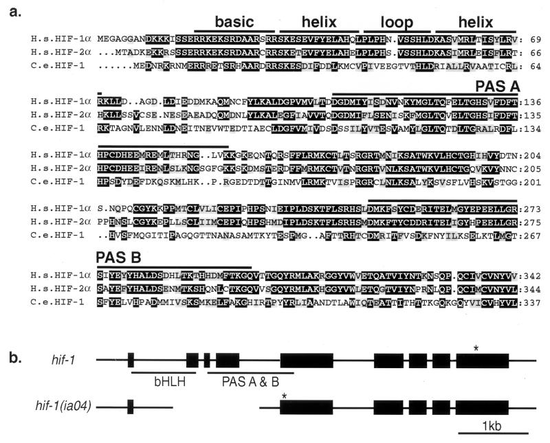
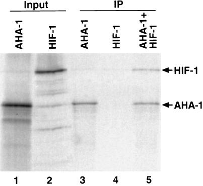
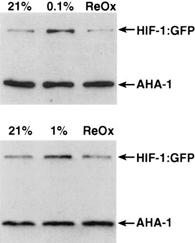
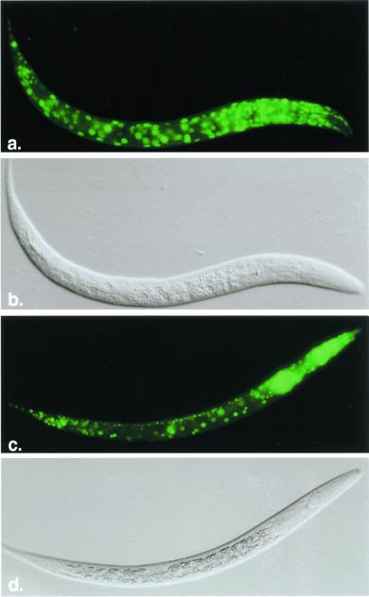
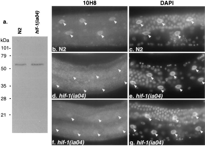
Hypoxia-inducible factor, a heterodimeric transcription complex, regulates cellular and systemic responses to low oxygen levels (hypoxia) during normal mammalian development or tumor progression. Here, we present evidence that a similar complex mediates response to hypoxia in Caenorhabditis elegans. This complex consists of HIF-1 and AHA-1, which are encoded by C. elegans homologs of the hypoxia-inducible factor (HIF) alpha and beta subunits, respectively. hif-1 mutants exhibit no severe defects under standard laboratory conditions, but they are unable to adapt to hypoxia. Although wild-type animals can survive and reproduce in 1% oxygen, the majority of hif-1-defective animals die in these conditions. We show that the expression of an HIF-1:green fluorescent protein fusion protein is induced by hypoxia and is subsequently reduced upon reoxygenation. Both hif-1 and aha-1 are expressed in most cell types, and the gene products can be coimmunoprecipitated. We conclude that the mechanisms of hypoxia signaling are likely conserved among metazoans. Additionally, we find that nuclear localization of AHA-1 is disrupted in an hif-1 mutant. This finding suggests that heterodimerization may be a prerequisite for efficient nuclear translocation of AHA-1.


Figures

Figure 1
Figure 1
(a) Conserved N-terminal domains of the predicted HIF-1 protein, and the Lif-1 genomic structure. Alignment of the N-terminal amino acid sequences of human HIF-1α (H.s. HIF-1α; U22431), HIF-2α (H.s. HIF-2α; U81983), and C. elegans HIF-1 (C.e. HIF-1; AF364604). Identical and similar amino acids are highlighted in black and gray, respectively. (b) Genomic structure of hif-1 and the hif-1 (ia04) deletion mutant. Boxes represent exons. * indicate the first stop codon in the hif-1 ORF. The ia04 mutation is a 1,231-bp deletion of the second, third, and fourth exons. This introduces a frameshift and premature stop in the mutant mRNA.
Figure 2
Figure 2
HIF-1 binds AHA-1. HIF-1 and AHA-1 were expressed in rabbit reticulocyte lysates in the presence of [35S]methionine (lanes 1 and 2), subjected to immunoprecipitation with an AHA-1-specific antibody (IP; lanes 3–5), and fractionated by SDS/PAGE. The antibody immunoprecipitates AHA-1 (lane 3) but not HIF-1 (lane 4). When AHA-1 and HIF-1 are coincubated, the antibody pulls down both proteins (lane 5). The molar ratio of HIF-1/AHA-1 in lane 5 is 1:2.5.
Figure 3
Figure 3
HIF-1:GFP protein levels are increased by hypoxia. The transgenic C. elegans strain pHJ06 Ex6, which expresses the complete HIF-1 protein fused to GFP, was harvested before (21% O2) and after incubation in 0.1% oxygen (Upper) or 1% oxygen (Lower) for 8 h. In each experiment, a fraction of the hypoxia-treated worms were reoxygenated for 10 min (ReOx). Worms were lysed by boiling in loading buffer, and equal amounts of total protein were loaded in each lane. The expression of HIF-1:GFP and AHA-1 was assayed by probing the immunoblot with a GFP-specific antibody and the AHA-1-specific mAb 10H8.
Figure 4
Figure 4
hif-1 and aha-1 promoters direct expression of GFP reporter genes in most cell types. Fluorescent images and Nomarski images of a C. elegans first-stage larvae expressing hif-1:gfp (a and b) and aha-1:gfp (c and d).
Figure 5
Figure 5
Subcellular localization of AHA-1 is disrupted in the hif-1 (ia04) mutant. (a) Equal amounts of total protein from wild-type (N2) or hif-1 (ia04) were size-fractionated by SDS/PAGE, blotted, and probed with the AHA-1-specific antibody 10H8. (b, d, and f) Immunostaining of intestinal cells with 10H8 in wild-type (N2) (b) or hif-1 mutant adults (d and f). (c, e, and g) The 4′,6-diamidino-2-phenylindole staining of the cells that are shown in b, d, and f, respectively. The arrowheads label the positions of intestinal cell nuclei.

References

    1. Guillemin K, Krasnow M A. Cell. 1997;89:9–12. - PubMed
    1. Semenza G L. Annu Rev Cell Dev Biol. 1999;15:551–578. - PubMed
    1. Wang G L, Jiang B H, Rue E A, Semenza G L. Proc Natl Acad Sci USA. 1995;92:5510–5514. - PMC - PubMed
    1. Iyer N V, Kotch L E, Agani F, Leung S W, Laughner E, Wenger R H, Gassmann M, Gearhart J D, Lawler A M, Yu A Y, Semenza G L. Genes Dev. 1998;12:149–162. - PMC - PubMed
    1. Ryan H E, Lo J, Johnson R S. EMBO J. 1998;17:3005–3015. - PMC - PubMed

Publication types

MeSH terms

Substances

Associated data

LinkOut - more resources