. 2001 Jul 3;98(14):7812-6.
doi: 10.1073/pnas.141235398. Epub 2001 Jun 26.

Id1 regulation of cellular senescence through transcriptional repression of p16/Ink4a

Affiliations

Id1 regulation of cellular senescence through transcriptional repression of p16/Ink4a

R M Alani et al. Proc Natl Acad Sci U S A. .

Abstract

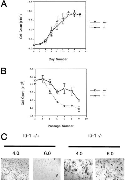
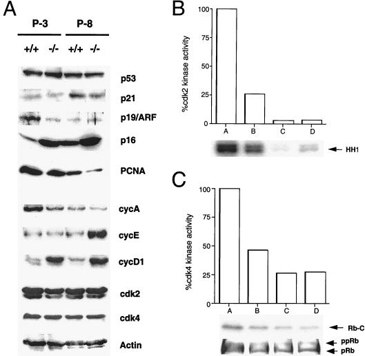
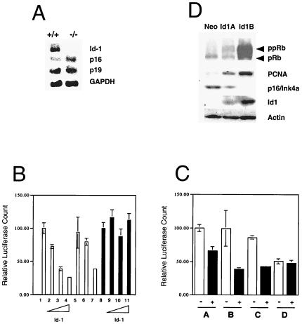
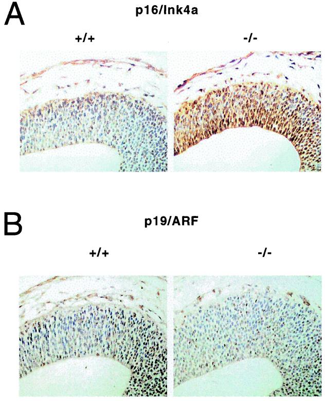
The Id family of helix-loop-helix (HLH) transcriptional regulatory proteins does not possess a basic DNA-binding domain and functions as a negative regulator of basic HLH transcription factors. Id proteins coordinate cell growth and differentiation pathways within mammalian cells and have been shown to regulate G(1)-S cell-cycle transitions. Although much recent data has implicated Id1 in playing a critical role in modulating cellular senescence, no direct genetic evidence has been reported to substantiate such work. Here we show that Id1-null primary mouse embryo fibroblasts undergo premature senescence despite normal growth profiles at early passage. These cells possess increased expression of the tumor-suppressor protein p16/Ink4a but not p19/ARF, and have decreased cyclin-dependent kinase (cdk) 2 and cdk4 kinase activity. We also show that Id1 is able to directly inhibit p16/Ink4a but not p19/ARF promoter activity via its HLH domain, and that Id1 inhibits transcriptional activation at E-boxes within the p16/Ink4a promoter. Our data provide, to our knowledge, the first genetic evidence for a role for Id1 as an inhibitor of cellular senescence and suggest that Id1 functions to delay cellular senescence through repression of p16/Ink4a. Because epigenetic and genetic abrogation of p16/Ink4a function has been implicated in the evolution of several human malignancies, we propose that transcriptional regulation of p16/Ink4a may also provide a mechanism for the dysregulation of normal cellular growth controls during the evolution of human malignancies.


Figures

Figure 1
Figure 1
Id1 −/− MEFs exhibit premature cellular senescence. (A) Growth curves for Id-1 +/+ and −/− MEFs at P-3. Replicate cultures of 1 × 10⁵ cells per 35-mm diameter dish were plated on day zero and cells were counted each day for 8 consecutive days (diao). (B) Senescence profile of Id-1 +/+ and −/− MEFs. P-3 MEFs were passaged on a 3T9 protocol in which 9 × 10⁵ cells were plated in a 60-mm dish and counted every 3 days with subsequent replating of 9 × 10⁵ cells at each passage until senescence was reached. (C) β-gal activity of P-8 MEFs. Cells were stained at pH 4.0 for lysosomal β-gal activity (positive control) and pH 6.0 for senescence-associated β-gal activity. Positive control is included to detail cellular morphology of Id1 +/+ vs. −/− cells at P-8 (×100).
Figure 2
Figure 2
Loss of Id1 affects the expression of cell-cycle regulatory proteins and cdk activity in early passage MEFs. (A) Western blot analysis of cell lysates from Id1 +/+ and −/− MEFs at early (P-3) and late (P-8) passages. Actin lane serves as loading control. (B and C) cdk2 and cdk4 kinase activity of Id1 +/+ and Id1 −/− MEFs. A, P-3 +/+ MEFs; B, P-3 −/− MEFs; C, P-8 +/+ MEFs; D, P-8 −/− MEFs. HH1 represents histone H1 phosphorylation and Rb-C represents phosphorylation of a C-terminal fragment of the retinoblastoma protein. Western blot for hyperphosphorylated (ppRb) or hypophosphorylated (pRb) is depicted below corresponding cdk4 kinase activity (29).
Figure 3
Figure 3
Id1 is a repressor of p16/Ink4a but not p19/ARF promoter activity. (A) Northern analysis of p16, p19, and Id1 expression at early (P-3) passage in Id1 wild-type (+/+) and Id1-null (−/−) MEFs. (B) Id1 repression of the p16/Ink4a promoter but not the p19/ARF promoter. 1, p16 promoter activity; 2–4, p16 promoter activity with 0.3, 1.5, or 3.0 μg of Id1; 5, p16 promoter + 3.0 μg of HLH dimerization domain of Id1 mutant of Id1; 6, p16 promoter + 3.0 μg of V91P mutant of Id1; 7, p16 promoter + 3.0 μg of ΔC-terminal mutant of Id1; 8, p19 promoter activity; 9–11, p19 promoter activity with 0.3, 1.5, or 3.0 μg of Id1. (C) Both the E-box at -349 and -615 bp of the p16/Ink4a promoter are critical for repression by Id1. White box, promoter alone; black box, promoter plus 1 μg of Id1; A, full-length p16 promoter (1.2 kb); B, p16 promoter with mutated E-box at -349 bp; C, p16 promoter with mutated E-box at -615 bp; D, p16 promoter with mutated E-boxes at both -349 and -615 bp. (D) Expression of p16 is repressed in Id1-transfected primary human keratinocytes. Western blot for expression of retinoblastoma protein, proliferating cell nuclear antigen, p16/Ink4a, and Id1 in transfected human foreskin keratinocytes expressing control plasmid (Neo) or Id1 for 1 month (Id1A) or 1 year (Id1B).
Figure 4
Figure 4
Expression p16 is up-regulated in the ventral telencephalon of Id1 −/− mouse embryos. Immunohistochemical analysis of E11.5 mouse embryo forebrains stained for p16/Ink4a (A) or p19/ARF (B) (brown). Blue represents counterstain.

References

    1. Massari M E, Murre C. Mol Cell Biol. 2000;20:429–440. - PMC - PubMed
    1. Norton J D. J Cell Sci. 2000;113:3897–3905. - PubMed
    1. Alani R M, Hasskarl J, Grace M, Hernandez M-C, Israel M A, Munger K. Proc Natl Acad Sci USA. 1999;96:9637–9641. - PMC - PubMed
    1. Nickoloff B J, Chaturvedi V, Bacon P, Qin J Z, Denning M F, Diaz M O. J Biol Chem. 2000;275:27501–27504. - PubMed
    1. Lundberg A S, Hahn W C, Gupta P, Weinberg R A. Curr Opin Cell Biol. 2000;12:705–709. - PubMed

Publication types

LinkOut - more resources