. 2001 Aug 31;276(35):33220-32.
doi: 10.1074/jbc.M104097200. Epub 2001 Jun 28.

The autocatalytic release of a putative RNA virus transcription factor from its polyprotein precursor involves two paralogous papain-like proteases that cleave the same peptide bond

Affiliations

The autocatalytic release of a putative RNA virus transcription factor from its polyprotein precursor involves two paralogous papain-like proteases that cleave the same peptide bond

J Ziebuhr et al. J Biol Chem. .

Abstract

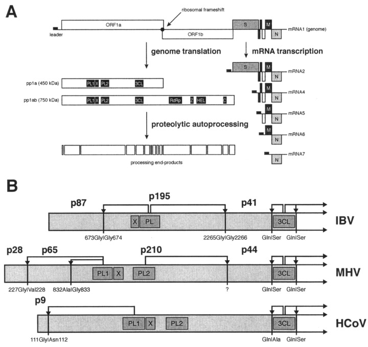
The largest replicative protein of coronaviruses is known as p195 in the avian infectious bronchitis virus (IBV) and p210 (p240) in the mouse hepatitis virus. It is autocatalytically released from the precursors pp1a and pp1ab by one zinc finger-containing papain-like protease (PLpro) in IBV and by two paralogous PLpros, PL1pro and PL2pro, in mouse hepatitis virus. The PLpro-containing proteins have been recently implicated in the control of coronavirus subgenomic mRNA synthesis (transcription). By using comparative sequence analysis, we now show that the respective proteins of all sequenced coronaviruses are flanked by two conserved PLpro cleavage sites and share a complex (multi)domain organization with PL1pro being inactivated in IBV. Based upon these predictions, the processing of the human coronavirus 229E p195/p210 N terminus was studied in detail. First, an 87-kDa protein (p87), which is derived from a pp1a/pp1ab region immediately upstream of p195/p210, was identified in human coronavirus 229E-infected cells. Second, in vitro synthesized proteins representing different parts of pp1a were autocatalytically processed at the predicted site. Surprisingly, both PL1pro and PL2pro cleaved between p87 and p195/p210. The PL1pro-mediated cleavage was slow and significantly suppressed by a non-proteolytic activity of PL2pro. In contrast, PL2pro, whose proteolytic activity and specificity were established in this study, cleaved the same site efficiently in the presence of the upstream domains. Third, a correlation was observed between the overlapping substrate specificities and the parallel evolution of PL1pro and PL2pro. Collectively, our results imply that the p195/p210 autoprocessing mechanisms may be conserved among coronaviruses to an extent not appreciated previously, with PL2pro playing a major role. A large subset of coronaviruses may employ two proteases to cleave the same site(s) and thus regulate the expression of the viral genome in a unique way.


Figures

Figure 1
Figure 1
Outline of theHCoVlife cycle and proteolytic processing of the N-terminal regions of coronavirus replicative polyproteins.A, ORFs in the polycistronic genome are indicated as boxes. The replicase gene, encompassing ORFs 1a and 1b, the gene for the surface glycoprotein protein, S, the triple-spanning membrane protein,M, and the nucleocapsid protein, N, are shown. The filled rectangle at the 5′ end of the genome represents the common leader sequence that is also present at the 5′ end of the subgenomic mRNAs that are shown below the genome. The conserved domains/functions encoded by the replicase gene are shown in the boxes depicting the two replicative polyproteins (pp1a and pp1ab). B, the N-terminal regions of the IBV, MHV, and HCoV replicative polyproteins pp1a/pp1ab are shown with the previously identified processing products and the corresponding cleavage sites (P1 and P1′ residues indicated). The following abbreviations are used:PL,papain-like protease; PL1, papain-like protease 1; X, domain conserved in coronaviruses, alphaviruses, rubiviruses, and hepatitis E virus (56); PL2, papain-like protease 2; 3CL, 3C-like protease;RdRp, RNA-dependent RNA polymerase;Z, putative zinc finger; HEL, NTPase/RNA helicase; C, conserved domain specific for nidoviruses (4).
Figure 2
Figure 2
Schematic representation of the N-terminalHCoVpp1a/pp1ab region and the proteins tested for proteolytic activity. The numbering of the amino acids is according to Ref.. The putative nucleophilic cysteine residues of PL1pro and PL2pro are indicated. The positions of the PL1pro p9↓p87 cleavage site (25) and two additional PLpro cleavage sites (this study) are given. The proteins tested for proteolytic activity are depicted on theleft, and the black lines designate these proteins in relation to their positions in pp1a and pp1ab. See “Experimental Procedures” for a description of the generation of each expression construct.
Figure 3
Figure 3
Multiple sequence alignment of the p195/p210 regions of coronavirus replicase polyproteins. An initial draft of this alignment was generated using the Dialign2 program (35) and subsequently improved with the ClustalX program (34). The alignment was further checked and corrected using results of a Macaw-mediated (36) analysis that involved all coronaviruses except MHVJ, which was excluded due to its closeness to MHVA. Five domains were recognized in the alignment, and their positions were indicated with ><. The borders of the domains are tentative. The alignments of the PL1pro and PL2pro regions were based on results of our previous analysis (32). For two regions that are located between domains X and PL2pro, and PL2pro and Y, respectively, no consistent alignments have been produced. Therefore, only the sizes of these regions are indicated. The pp1a position of the rightmost residue in an alignment row is indicated at the right side. The shading of individual residues in the alignment was done according to a four-level conservation; black background and white letters, gray background andwhite letters, gray background and black letters, respectively, indicate residues that are conserved in 100, 80, and 60% of the sequences. Groups of conserved amino acids are as follows: IVLM; FYW; KRH; DNQE; ST; AG. According to the Macaw, four blocks, which are labeled with letters from A to D abovethe alignment and are discussed in the main text are statistically significant for the entire pp1a searching space: A, p = 1.1e−002; B, p = 2.1e−002; C, p = 4.2e−006; and D, p = 3.9e−015. Two hydrophobic regions predicted to be trans-membrane domains (40) are marked withdashed lines and denoted with TM1 and TM2, respectively. Other highlights are as follows: +, catalytic Cys and His residues of PLpros; #, postulated metal-chelating Cys and His residues of the PLpro zinc fingers; @, conserved Cys and His residues of domain Y; ‖, cleavage sites of PLpros. MHVA and MHVJ, MHV strains A59 and JHM. The National Center for Biotechnology Information sequence ID: IBV, 138147; HCoV, 464694; TGEV, 872319; MHVA, 453423 (nucleotide); MHVJ, 266958 (corrected according to Ref. 57); all sequences are for proteins unless otherwise specified.
Figure 4
Figure 4
Detection of an ORF1a-encoded 87-kDa cleavage product in HCoV-infected cells. Metabolically labeled lysates from 3 × 10⁵ mock-infected (M) (lanes 1 and 3) or HCoV-infected (I) (lanes 2 and 4) MRC-5 cells were analyzed by SDS-polyacrylamide gel electrophoresis in a 10–17% acrylamide gradient gel after immunoprecipitation with α-H2 antiserum (lanes 3 and 4) or the corresponding preimmune serum (lanes 1 and 2). Antiserum α-H2 recognizes the HCoV ORF1a-encoded amino acids 112–322. The cells were labeled from 7 to 9.5-h postinfection with 100 µCi of [35S]methionine per ml. Sizes of molecular mass markers (CFA 626; Amersham Pharmacia Biotech) with masses in kilodaltons as well as the 230- and 87-kDa processing products, p230 and p87, respectively, are indicated.
Figure 5
Figure 5
Evidence for a second PL1pro cleavage site in theHCoVpp1a and pp1ab polyproteins.A, in vitro translation reactions of capped RNAs derived fromEcoRI-linearized plasmids pBST-111–103 and pBST-111–103_C1054A. The respective RNAs encode the pp1a/1ab amino acids 717–1285 (WT, lanes 1 and 2) or the same sequence with an active-site replacement of the catalytic Cys1054 (C1054A, lanes 3 and 4). The translation reactions were done as described under “Experimental Procedures,” and the reaction products were either analyzed directly (lanes 1 and 3) or after further incubation for 120 min (lanes 2 and 4). The positions of full-length precursor proteins and cleavage products are indicated.B, a protein called pp717–1285_VM was translated in a reticulocyte lysate in the presence of [35S]methionine. Except for three amino acid substitutions (V900M, V906M, and V908M), which had been introduced downstream to the presumed cleavage site, this protein contained the HCoV pp1a/1ab wild-type sequence from residues 717 to 1285. The translation reaction was incubated for 160 min at 30 °C, and the reaction products were separated on an SDS-12.5% polyacrylamide gel. After electrophoretic transfer to PVDF membranes, the position of the C-terminal cleavage product was determined by autoradiography. The isolated protein was subjected to 16 cycles of Edman degradation, and the distribution of radiolabeled amino acids was determined by scintillation counting. The amino acid sequence of pp1a and pp1ab from positions 895 to 913 is shown. The amino acids Met900, Met906, and Met908 present in pp717–1285_VM are shown in boldface type, and the newly identified PL1pro cleavage site is indicated by an arrow.
Figure 7
Figure 7
Relationship between PL1pro and PL2pro proteolytic activities.A, in vitrotranslation reactions of capped RNAs encoding the pp1a/1ab amino acids 717–1285 (lanes 1 and 2), amino acids 717–1436 (lanes 3 and 4), amino acids 717–1910 (lanes 5 and 6), and amino acids 759–1910 (lanes 7 and 8). The proteins to be tested for proteolytic activity were translated in rabbit reticulocyte lysates in the presence of [35S]methionine at 30 °C for 40 min. After the termination of translation, the reaction products were either analyzed directly (lanes 1, 3, 5, and 7) or after further incubation for 120 min (lanes 2, 4, 6, and8). The analysis was done by SDS-polyacrylamide gel electrophoresis in a 10–17% polyacrylamide gradient gel. Full-length precursor proteins and major processing products are indicated (*, precursor protein; ●, processing product). Also, the calculated cleavage activities of the full-length precursor proteins are given (see “Experimental Procedures” for details). B,proteolytic activities of pp717–1910-derived proteins carrying active-site mutations in the two HCoV PLpro domains. The proteins to be tested for proteolytic activity were translated in rabbit reticulocyte lysates in the presence of [35S]methionine at 30 °C for 40 min. After termination of translation, the reaction products were separated by SDS-polyacrylamide gel electrophoresis. They were either analyzed directly (lanes 1, 3, 5, 7, and9) or after further incubation for 120 min (lanes 2, 4, 6, 8, and 10). The proteins, which all encompassed the HCoV pp1a/pp1ab amino acids 717–1910, contained Cys-to-Ala replacements of the putative nucleophilic residues of PL1pro (C1054A,lanes 1 and 2) and PL2pro (C1701A, lanes 5 and 6), 8-amino acid deletions including the putative nucleophilic residues of PL1pro (Δ1054–1061, lanes 3 and4) and PL2pro (Δ1701–1708, lanes 7 and8) or wild-type sequence (lanes 9and10). The positions of full-length precursor proteins and cleavage products are indicated. Also the calculated cleavage activities of the full-length precursor proteins are given.
Figure 6
Figure 6
Proteolytic activity of the PL2pro domain and identification of its cleavage site.A, the HCoV ORF1a-encoded amino acids 717–1910 were translated in rabbit reticulocyte lysates in the presence of [35S]methionine. The proteins to be tested for proteolytic activity contained wild-type sequence (lanes 1 and 2) or the same sequence with active-site replacements in PL1pro (C1054A, lanes 3 and4) and both PL1pro and PL2pro (C1054A and W1702L), respectively. The proteins were translated at 30 °C for 40 min, and after the termination of translation, the reaction products were either analyzed directly (lanes 1, 3,and 5) or after further incubation for 120 min (lanes 2, 4,and6). The analysis was done by SDS-polyacrylamide gel electrophoresis in a 10–17% polyacrylamide gradient gel. The positions of full-length precursor proteins and cleavage products are indicated. B, a protein called pp717–1910_C1054A-VM was translated in a reticulocyte lysate in the presence of [35S]methionine. Except for a PL1pro-inactivating amino acid replacement (C1054A) and three additional substitutions (V900M, V906M, and V908M), which had been introduced downstream to the predicted cleavage site, this protein contained the HCoV pp1a/1ab wild-type sequence from residues 717 to 1910. The translation reaction was incubated for 160 min at 30 °C, and the reaction products were separated on an SDS-10% polyacrylamide gel. After electro- phoretic transfer to PVDF membranes, the position of the C-terminal cleavage product was determined by autoradiography. The isolated protein was subjected to 16 cycles of Edman degradation, and the distribution of radiolabeled amino acids was determined by scintillation counting. The amino acid sequence of pp1a and pp1ab from positions 895 to 913 is shown. The amino acids Met900, Met906, and Met908 present in pp717–1910_C1054A-VM are shown in boldface type, and the deduced PL2pro cleavage site is indicated by an arrow.
Figure 8
Figure 8
Phylogenetic relationships among coronavirus PLpros.A, expected, and B,reconstructed and unrooted trees for coronavirus PLpros. The genetic groups recognized in coronaviruses are uniquely colored. A,the topology of this tree of PLpros was approximated on the basis of sequence comparisons of other coronavirus replicative enzymes3 that result in a tree topology identical to the one shown here for the PL1pro and PL2pro subgroups. This representation is called the “expected” tree. The two lineages that were reshuffled in B, MHVA/BCoVL_PL2 and TGEV/HCoV_PL1, are shown with a gray background. B, the “reconstructed” tree was generated using an alignment of PLpros (Fig. 3; see also Fig. 2A in (32)) and the NJ algorithm with the Kimura correction as implemented in the ClustalX program. The alignment included the following sequences: HCoV_PL1pro (1043–1227), HCoV_PL2pro (1690–1885), TGEV_PL1pro (1082–1266), TGEV_PL2pro (1577–1763), MHVA_PL1 (1110–1292), MHVA_PL2 (1705–1896), BCoVL_PL1 (1063–1245), BCoVL_PL2 (1660–1851) and IBV_PL2 (1263–1457). BCoVL is from Footnote 3; for the sources of the other sequences see Fig. 3. The number of trees, in which a particular bifurcation was sustained in the course of 1000 bootstrap simulations, is given at each node.
Figure 9
Figure 9
Proposed scheme for the proteolytic processing of coronavirus replicative polyproteins by the accessory proteases PL1pro and PL2pro. Cleavage sites (P1 and P1′ residues indicated) identified in the pp1a/pp1ab proteins of IBV, MHV, HCoV, and TGEV and the corresponding processing products identified in virus-infected cells are shown. Putative cleavage sites, which are predicted on the basis of the results of the present study, are indicated by ?. Also, the protease domains responsible for specific cleavages are given, with solid lines indicating experimentally characterized cleavages and dotted linesindicating predicted cleavages. The proteolytically inactive IBV PL1pro is marked by a black background color. For other abbreviations see Figs. 1B and 3.

References

    1. Kräusslich H.G., Wimmer E. Annu. Rev. Biochem. 1988;57:701–754. - PubMed
    1. Dougherty W.G., Semler B.L. Microbiol. Rev. 1993;57:781–822. - PMC - PubMed
    1. Gorbalenya A.E., Snijder E.J. Perspect. Drug Discov. Des. 1996;6:64–86. - PMC - PubMed
    1. den Boon J.A., Snijder E.J., Chirnside E.D., de Vries A.A., Horzinek M.C., Spaan W.J. J. Virol. 1991;65:2910–2920. - PMC - PubMed
    1. Cavanagh D. Arch. Virol. 1997;142:629–633. - PubMed

Publication types

MeSH terms

LinkOut - more resources