Review
. 2001 Jul;14(3):447-75.
doi: 10.1128/CMR.14.3.447-475.2001.

Biology of Giardia lamblia

Affiliations
Review

Biology of Giardia lamblia

R D Adam. Clin Microbiol Rev. 2001 Jul.

Abstract

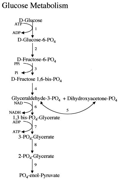
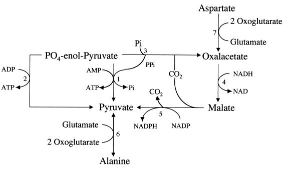
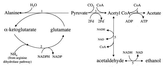
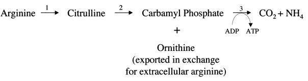
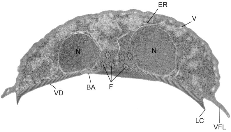
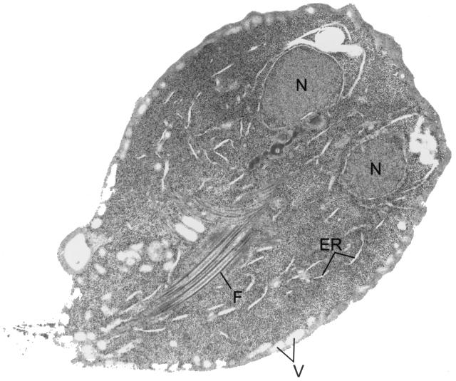
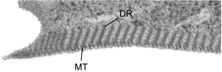
Giardia lamblia is a common cause of diarrhea in humans and other mammals throughout the world. It can be distinguished from other Giardia species by light or electron microscopy. The two major genotypes of G. lamblia that infect humans are so different genetically and biologically that they may warrant separate species or subspecies designations. Trophozoites have nuclei and a well-developed cytoskeleton but lack mitochondria, peroxisomes, and the components of oxidative phosphorylation. They have an endomembrane system with at least some characteristics of the Golgi complex and encoplasmic reticulum, which becomes more extensive in encysting organisms. The primitive nature of the organelles and metabolism, as well as small-subunit rRNA phylogeny, has led to the proposal that Giardia spp. are among the most primitive eukaryotes. G. lamblia probably has a ploidy of 4 and a genome size of approximately 10 to 12 Mb divided among five chromosomes. Most genes have short 5' and 3' untranslated regions and promoter regions that are near the initiation codon. Trophozoites exhibit antigenic variation of an extensive repertoire of cysteine-rich variant-specific surface proteins. Expression is allele specific, and changes in expression from one vsp gene to another have not been associated with sequence alterations or gene rearrangements. The Giardia genome project promises to greatly increase our understanding of this interesting and enigmatic organism.


Figures

FIG. 1
FIG. 1
Metabolism of glucose to phosphoenolpyruvate. Many of the enzymes have been documented in terms of enzymatic activity, isolation of the enzyme, or cloning of the gene encoding the enzyme. The enzymes are labeled as follows: 1, hexokinase (188); 2, glucose phosphate isomerase (proposed); 3, pyrophosphate-dependent phosphofructokinase (186, 219, 274, 289); 4, fructose bisphosphate aldolase (128, 188); 5, triosephosphate isomerase (238); 6, glyceraldehyde-3-phosphate dehydrogenase (287); 7, phosphoglycerate kinase (proposed); 8, phosphoglyceromutase (proposed); 9, enolase (proposed). Certain enzymes (for steps , , , and 9) are suggested on the basis of pathways in other organisms but have not yet been proved for Giardia. This figure has been adapted from material presented in prior reviews (55, 146, 150) and updated from more recent literature as cited for individual enzymes.
FIG. 2
FIG. 2
Intermediary pathways to synthesis of pyruvate. 1, Pyruvate phosphate dikinase (132, 141); 2, pyruvate kinase (271) (the relative roles of pyruvate phosphate dikinase and pyruvate kinase in the conversion of phosphoenolpyruvate to pyruvate have not been determined); 3, phosphoenolpyruvate carboxyphosphotransferase; 4, malate dehydrogenase (188, 285); 5, malate dehydrogenase (decarboxylating) (188, 294); 6, alanine transaminase (72, 263); 7, aspartate transaminase (215). Pi, inorganic phosphate. This figure has been adapted from material presented in prior reviews (55, 215) and updated from more recent literature (see citations for individual enzymes).
FIG. 3
FIG. 3
End product synthesis from pyruvate. The enzymes are labeled as follows: 1, alanine aminotransferase (72, 263) (alanine is produced only under anaerobic conditions [265]); 2, GDH (263, 272, 359); 3, PFOR (327) (ferredoxin rather than NAD as the electron acceptor [326]); 4, acetyl-CoA synthetase (ADP forming) (293); 5, alcohol dehydrogenase E (ADHE) (has acetaldehyde dehydrogenase activity in the amino terminus which catalyzes the conversion of acetyl-CoA to acetaldehyde and alcohol dehydrogenase activity in the carboxy terminus which catalyzes the conversion of acetaldehyde to ethanol) (59, 292). Acetate is the major product under aerobic conditions; ethanol and alanine are preferentially produced under anaerobic conditions (265).
FIG. 4
FIG. 4
Arginine dihydrolase pathway (74, 297, 300). The enzymes are labeled as follows: 1, arginine deiminase (163); 2, ornithine transcarbamoylase (297, 300); 3, carbamate kinase (226).
FIG. 5
FIG. 5
Purine ribonucleoside salvage pathways. The enzymes are labeled as follows: 1, adenosine hydrolase (339); 2, adenine phosphoribosyltransferase (APRTase); (339); 3, guanosine hydrolase (339); 4, guanine phosphoribosyltransferase (GPRTase) (339).
FIG. 6
FIG. 6
Pyrimidine ribonucleoside salvage pathways. The enzymes are labeled as follows: 1, uracil phosphoribosyltransferase (UPRTase) (58); 2, uridine/thymine phosphorylase (181) (a uridine hydrolase [13] has not been confirmed); 3, uridine phosphotransferase (kinase) (13) (not confirmed [336]); 4, UMP kinase (151); 5, UDP kinase (151); 6, CTP synthetase (151, 187); 7, CDP kinase (151); 8, cytosine phosphoribosyltransferase (CPRTase) (13) (low level of activity [151]); 9, cytidine hydrolase (13); 10, cytidine deaminase (336). The initial pyrimidine salvage pathway was described in (reference) and has been updated from more recent literature (see the references cited for individual enzymes).
FIG. 7
FIG. 7
Deoxynucleoside salvage pathways. The enzymes are labeled as follows: 1, pyrimidine deoxynucleoside kinase (176); 2, purine deoxynucleoside kinase (176).
FIG. 8
FIG. 8
Trophozoite coronal section. A coronal view of a trophozoite demonstrates the nuclei (N), endoplasmic reticulum (ER), flagella (F), and vacuoles (V). A mechanical suction is formed when the ventral disk (VD) attaches to an intestinal or glass surface. Components of the ventral disk include the bare area (BA), lateral crest (LC), and ventrolateral flange (VLF). A magnified view of the ventral disk is shown in Fig. 10.
FIG. 9
FIG. 9
Trophozoite cross section. A cross-sectional view of a trophozoite demonstrates the nuclei (N), flagella (F), vacuoles (V), and endoplasmic reticulum (ER).
FIG. 10
FIG. 10
Close-up of the ventral disk. A magnified view of the ventral disk shows the microtubules (MT) and microribbons or dorsal ribbons (DR).
FIG. 11
FIG. 11
Enzymatic pathway for the synthesis of N-acetylgalactosamine, the major carbohydrate portion of the cyst wall. Activities for all these enzymes have been shown in reference (203) in a mixture of vegetative and encysting trophozoites, and a proposed pathway was presented. The enzymes are labeled as follows: 1, glucosamine-6-phosphate-isomerase (164, 312, 334); 2, glucosamine-6-phosphate N-acetylase; 3, phosphoacetylglucosamine mutase; 4, UDP-N-acetylglucosamine pyrophosphorylase (40, 41); 5, UDP-N-acetylglucosamine 4′ epimerase.
FIG. 12
FIG. 12
PFGE separation of JH and ISR. A PFGE separation of JH (genotype A-2) chromosomes demonstrates five distinct bands, while a PFGE separation of ISR (genotype A-1) chromosomes demonstrates four intensely staining and two more faintly staining bands. A series of chromosome-specific probes have been used to identify five distinct linkage groups corresponding to the five bands of the JH isolate (7, 177). The fainter bands of the ISR isolate represent size variants of chromosome 1, as demonstrated by a set of chromosome-specific probes (7) as well as by detailed mapping of chromosome 1 (139). One of the size variants comigrates with chromosome 2; hence that band is labeled 1b,2. Part of the size variation of chromosome 1 is due to variation in the number of telomeric rDNA repeats (4), while much of the remainder is due to variation near the other telomere (139).
FIG. 13
FIG. 13
Southern blot demonstrating allelic heterozygosity of the vspC5 gene as well as members of the family of genes related to vspC5. Reproduced from reference with permission of Elsevier Science. Genomic DNA from WBA6 (expressing VSPA6), WB1269 (an antigenic variant derived from WBA6 and expressing VSP1269 [CRP72]), and WBC5 (an antigenic variant derived from WB1269 and expressing VSPC5) was digested with BamHI, blotted, and probed. (A) Results of moderate-stringency washing after hybridization with BS176, a probe extending from −93 to +83 of vspC5. (B) High-stringency washing. (C) Results after hybridization with the vspC5 105-bp repeat. The bands labeled C5.1 to C5.4 indicate four alleles of the vspC5 gene. Cosmids were cloned for each of the four alleles and were all identical in the regions flanking the coding region. These cosmids were mapped to a single chromosomal location by using PFGE and rare-cutting restriction enzymes, demonstrating that these genes were truly allelic in nature. The largest of the four bands probably represents the expressed allele as assessed by the size of the transcript on a Northern blot (358). Note that the smallest of the four alleles is deleted from the WBC5 genome. The mechanism for this deletion has not been determined. The bands labeled S1 to S4 represent four different vsp genes (vspC5-S1 to vspC5-S4) that are similar to vspC5 in the 5′ region. Cosmids for each of these genes were cloned; each was different in the flanking regions, and each mapped to a different chromosomal location by PFGE. The sequences of vspC5-S1 and vspC5-S2 were >96% identical to that of vspC5 in the region of the 176-bp probe. The disappearance of the vspC5-S3 band after high-stringency washing (B) indicates that it has less similarity to vspC5. The bands for vspC5-S1 to vspC5-S4 are not seen with the repeat probe (C), consistent with the lack of the 105-bp repeat in these genes.

References

    1. Reference deleted.
    1. Abaza S M, Sullivan J J, Visvesvara G S. Isoenzyme profiles of four strains of Giardia lamblia and their infectivity to jirds. Am J Trop Med Hyg. 1991;44:63–68. - PubMed
    1. Adam R D. The biology of Giardia spp. Microbiol Rev. 1991;55:706–732. - PMC - PubMed
    1. Adam R D. Chromosome-size variation in Giardia lamblia: the role of rDNA repeats. Nucleic Acids Res. 1992;20:3057–3061. - PMC - PubMed
    1. Adam R D. The Giardia lamblia genome. Int J Parasitol. 2000;30:475–484. - PubMed

Substances