Review
. 2001 Jul;14(3):584-640.
doi: 10.1128/CMR.14.3.584-640.2001.

Listeria pathogenesis and molecular virulence determinants

Affiliations
Review

Listeria pathogenesis and molecular virulence determinants

J A Vázquez-Boland et al. Clin Microbiol Rev. 2001 Jul.

Abstract

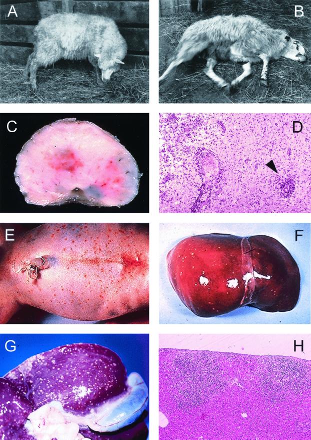
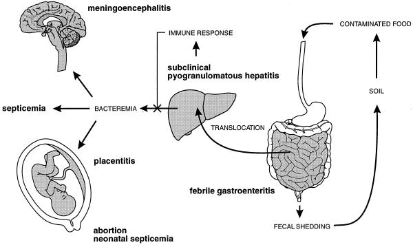
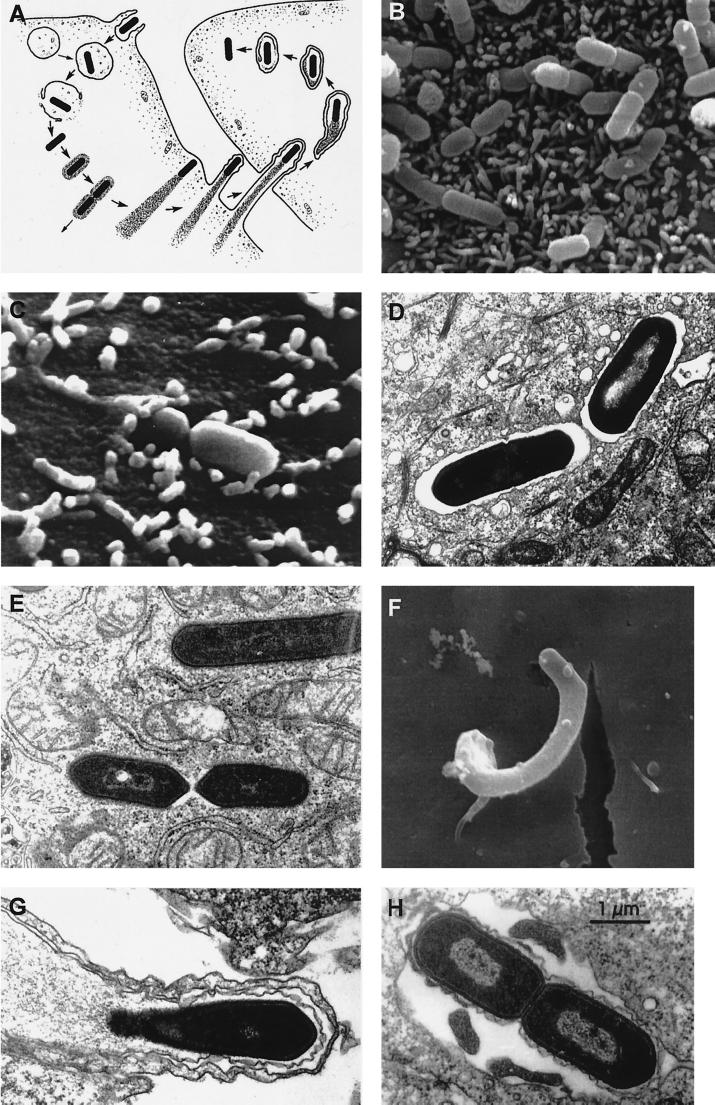
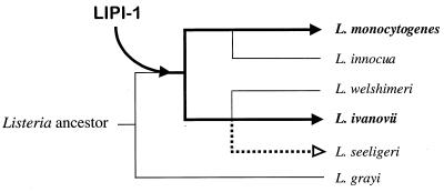
The gram-positive bacterium Listeria monocytogenes is the causative agent of listeriosis, a highly fatal opportunistic foodborne infection. Pregnant women, neonates, the elderly, and debilitated or immunocompromised patients in general are predominantly affected, although the disease can also develop in normal individuals. Clinical manifestations of invasive listeriosis are usually severe and include abortion, sepsis, and meningoencephalitis. Listeriosis can also manifest as a febrile gastroenteritis syndrome. In addition to humans, L. monocytogenes affects many vertebrate species, including birds. Listeria ivanovii, a second pathogenic species of the genus, is specific for ruminants. Our current view of the pathophysiology of listeriosis derives largely from studies with the mouse infection model. Pathogenic listeriae enter the host primarily through the intestine. The liver is thought to be their first target organ after intestinal translocation. In the liver, listeriae actively multiply until the infection is controlled by a cell-mediated immune response. This initial, subclinical step of listeriosis is thought to be common due to the frequent presence of pathogenic L. monocytogenes in food. In normal individuals, the continual exposure to listerial antigens probably contributes to the maintenance of anti-Listeria memory T cells. However, in debilitated and immunocompromised patients, the unrestricted proliferation of listeriae in the liver may result in prolonged low-level bacteremia, leading to invasion of the preferred secondary target organs (the brain and the gravid uterus) and to overt clinical disease. L. monocytogenes and L. ivanovii are facultative intracellular parasites able to survive in macrophages and to invade a variety of normally nonphagocytic cells, such as epithelial cells, hepatocytes, and endothelial cells. In all these cell types, pathogenic listeriae go through an intracellular life cycle involving early escape from the phagocytic vacuole, rapid intracytoplasmic multiplication, bacterially induced actin-based motility, and direct spread to neighboring cells, in which they reinitiate the cycle. In this way, listeriae disseminate in host tissues sheltered from the humoral arm of the immune system. Over the last 15 years, a number of virulence factors involved in key steps of this intracellular life cycle have been identified. This review describes in detail the molecular determinants of Listeria virulence and their mechanism of action and summarizes the current knowledge on the pathophysiology of listeriosis and the cell biology and host cell responses to Listeria infection. This article provides an updated perspective of the development of our understanding of Listeria pathogenesis from the first molecular genetic analyses of virulence mechanisms reported in 1985 until the start of the genomic era of Listeria research.


Figures

FIG. 1
FIG. 1
Clinical and pathological characteristics of Listeria infection in animals and humans. (A to D) Neuromeningeal listeriosis in sheep. (A) Typical aspect of circling disease, first described by Gill in 1933 in New Zealand (215, 216) and the most characteristic clinical manifestation of listeriosis in ruminants; the syndrome is characterized by involuntary torticollis and walking aimlessly in circles as a result of brainstem lesions. (B) In a further step of the infectious process, animals lie on the ground with evident signs of uncoordination (paddling movements) and cranial nerve paralysis (strabismus, salivation, etc.). (C) Section of the medulla oblongata of a sheep with listerial rhombencephalitis, showing clear inflammatory lesions in the brain tissue. (D) Microscopic preparation of the brainstem showing extensive parenchymal inflammatory infiltration and typical perivascular cuffing (one is indicated by an arrowhead) (panels C and D copyright M. Domingo, Barcelona, Spain). (E and F) Stillborn with generalized L. monocytogenes infection, a clinical condition also known as granulomatosis infantiseptica and characterized by the presence of military-disseminated pyogranulomatous lesions on the body surface (E) and internal organs (F, liver of the fetus in E) and a high mortality. (G and H) Extensive pyogranulomatous hepatitis in a lamb experimentally infected with L. ivanovii, with multiple necrotic foci in the liver surface (G) and the parenchyma (H, hematoxylin/eosin-stained cut from the liver in G, showing two subcapsular pyogranulomes [magnification, ×60]).
FIG. 2
FIG. 2
Schematic representation of the pathophysiology of Listeria infection. See text for details.
FIG. 3
FIG. 3
Stages of listerial intracellular parasitism. (A) Scheme of the intracellular life cycle of pathogenic Listeria spp. (reproduced from reference with permission of the Rockefeller University Press). In anticlockwise order: entry into the host cell by induced phagocytosis, transient residence within a phagocytic vacuole, escape from the phagosome into the cytoplasm, cytosolic replication and recruitment of host cell actin onto the bacterial surface, actin-based motility, formation of pseudopods, phagocytosis of the pseudopods by neighboring cells, formation of a double-membrane phagosome, escape from this secondary phagosome, and reinitiation of the cycle. (B to H) Scanning and transmission electron micrographs of cell monolayers infected with L. monocytogenes (B) Numerous bacteria adhering to the microvilli of a Caco-2 cell (30 min after infection). (C) Two bacteria in the process of invasion (Caco-2 cell, 30 min postinfection). (D) Two intracellular bacteria soon after phagocytosis, still surrounded by the membranes of the phagocytic vacuole (Caco-2 cell, 1 h postinfection). (E) Intracellular Listeria cells free in the host cell cytoplasm after escape from the phagosome (Caco-2 cell, 2 h postinfection). (F) Pseudopod-like membrane protrusion induced by moving Listeria cells, with the bacterium being evident at the tip (brain microvascular endothelial cell, 4 h postinfection; taken from reference with permission). (G) Section of a pseudopod-like structure in which a thin cytoplasmic extension of an infected cell is protruding into a neighboring noninfected cell (notice that the protrusion is covered by two membranes) (Caco-2 cell, 4 h postinfection). (H) Bacteria in a double-membrane vacuole formed during cell-to-cell spread (Caco-2 cell, 4 h postinfection).
FIG. 4
FIG. 4
Pore-forming activity of LLO. Sheep erythrocyte ghost after treatment with purified LLO, showing the ring-shaped oligomeric structures of the toxin attached to the membrane. (Bar, 100 nm.) (Reproduced from reference with permission of the publisher).
FIG. 5
FIG. 5
Hemolytic activities of pathogenic Listeria spp. Left panels, spontaneous hemolysis of L. monocytogenes (L. mo.) and L. ivanovii (L. iv.) on sheep blood agar; compare the narrow ring of β-hemolysis produced by L. monocytogenes (here exceptionally evident) with the striking multizonal lytic activity of L. ivanovii, which is due to the production of an additional cytolytic factor (the sphingomyelinase SmcL). Bacteria were cultured at 37°C for 36 h and then left at 4°C for 48 h. Right panels, synergistic (CAMP-like) hemolysis of L. ivanovii and β-toxin (sphingomyelinase)-producing S. aureus (S. au.) with R. equi (vertical streak); L. ivanovii and S. aureus elicit identical shovel-shaped patches of synergistic hemolysis, reflecting functional relatedness of the sphingomyelinases C produced by these two bacteria (see text and reference for details). The CAMP-like effect results from the total lysis of the halo of sphingomyelinase-damaged erythrocytes exposed to a cholesterol oxidase from R. equi (467). As shown in the two upper right panels, the L. ivanovii sphingomyelinase is active towards sheep erythrocytes (which contain 51% sphingomyelin in the membrane) but not towards horse erythrocytes (13.5% sphingomyelin only). Note that the inner zone of spontaneous hemolysis that surrounds L. ivanovii colonies, due to the activity of the hemolysin (ivanolysin O), is not influenced by the source of erythrocytes. Bacteria were cultured at 37°C for 24 h.
FIG. 6
FIG. 6
Actin-based intracellular motility. (A) Schematic structure and functional motifs of ActA (see text for details; amino acids numbered according to references and 672). (B) Cos-1 cells infected with L. monocytogenes and labeled with FITC-phalloidin (actin stain, green) and rhodamine-conjugated anti-Listeria antibody (red) at 4 h postinfection; moving bacteria with actin tails are visible. (C and D) Giemsa stain of Caco-2 cells infected with L. monocytogenes wild-type (C) and ΔactA mutant (D) at 4 h postinfection; ΔactA mutant bacteria do not move intracellularly and do not spread to neighboring cells, growing in the cytoplasm as a microcolony close to the nucleus.
FIG. 6
FIG. 6
Actin-based intracellular motility. (A) Schematic structure and functional motifs of ActA (see text for details; amino acids numbered according to references and 672). (B) Cos-1 cells infected with L. monocytogenes and labeled with FITC-phalloidin (actin stain, green) and rhodamine-conjugated anti-Listeria antibody (red) at 4 h postinfection; moving bacteria with actin tails are visible. (C and D) Giemsa stain of Caco-2 cells infected with L. monocytogenes wild-type (C) and ΔactA mutant (D) at 4 h postinfection; ΔactA mutant bacteria do not move intracellularly and do not spread to neighboring cells, growing in the cytoplasm as a microcolony close to the nucleus.
FIG. 7
FIG. 7
Structure of the members of the internalin multigene family from L. monocytogenes and L. ivanovii (reproduced with modifications from reference with permission of the publisher with data from Dominguez-Bernal et al. [unpublished data]). S, signal peptide; B, B-repeats; C, Csa domain repeats C-repeat; D, D-repeats. See text for details.
FIG. 8
FIG. 8
Physical and transcriptional organization of the central virulence gene cluster (LIPI-1) of L. monocytogenes and structure of the locus in other Listeria spp. Genes belonging to LIPI-1 are in grey, and the flanking loci are in black. Dotted lines above L. monocytogenes LIPI-1 genes indicate known transcripts. LIPI-1 is inserted in a chromosomal region delimited by the prs and ldh genes, encoding the housekeeping enzymes phosphoribosyl-pyrophosphate synthase and lactate dehydrogenase, respectively. In the plcB-ldh intergenic region, two ORFs, orfA and orfB, are found in all Listeria spp., indicating that the insertion point of LIPI-1 is between the prs and orfB loci. In the plcB-orfB intergenic region of L. monocytogenes, there are two small ORFs, orfX and orfZ (stippled), which delimit the putative excision point of LIPI-1 in L. innocua. In the plcB-orfB intergenic region of L. ivanovii, two small ORFs, orfX (encoding a homolog of the orfX product from L. monocytogenes) and orfL (stippled), are also present, which, like those in L. monocytogenes, delimit the deletion point of LIPI-1 in the nonpathogenic species L. welshimeri. orfZ and orfL encode polypeptides with significant similarity to bacteriophage proteins (respectively Orf6 of the A511 Listeria phage and a serine/threonine protein phosphatase from λ [88a, 670a]), suggesting that LIPI-1 was originally acquired by phage transduction (see text for details). The additional ORFs found in the L. seeligeri virulence gene cluster are hatched (the orfK product shows 39% similarity with the phosphatidylinositol phospholipase PlcA and dplcB is a truncated duplicate of the plcB gene). The figure has been constructed using data from references , , , and and unpublished results from J.K. (L. seeligeri) and B.G.-Z. and J.-A.V.-B. (L. ivanovii). (Adapted from reference with permission of the publisher.)
FIG. 9
FIG. 9
Evolution of the central virulence gene cluster (LIPI-1) in the genus Listeria. The presence of LIPI-1 at the same chromosomal position in various species belonging to distinct evolutionary branches of the genus suggests that the virulence gene cluster was acquired before speciation (see text for details). Thick lines indicate the evolutionary pathway followed by LIPI-1; solid arrows indicate conservation of a functional LIPI-1 (in L. monocytogenes and L. ivanovii), whereas the dotted lines with an open arrowhead indicate functional corruption of the virulence gene cluster (in L. seeligeri). L. innocua and L. welshimeri presumably arose by excision of LIPI-1 in the corresponding evolutionary branches. The dendrogram is a schematic reconstruction of the phylogeny of Listeria according to references and . (Adapted from reference with permission of the publisher.)
FIG. 10
FIG. 10
Internalin islands and islets of L. monocytogenes and L. ivanovii. The scheme shows the different gene arrangements found for the same internalin locus in two isolates of L. monocytogenes, possibly resulting from rearrangements between inl genes (inlH presumably arose by recombination of the 5′-terminal part of inlC2 and the 3′-terminal part of inlD [350, 527]). See text for details.
FIG. 11
FIG. 11
Structure of PrfA (A) and its target DNA sequences in PrfA-regulated promoters (B). See text for details. In PrfA, amino acid coordinates correspond to the peptide sequence deduced from the prfA gene (position 1 corresponds to the Met residue of the first triplet), whereas in Crp they correspond to the actual amino acid position in the primary structure of the native protein, which lacks the residue encoded by the first triplet. Therefore, the amino acid numbering of Crp is shifted one position with respect to that of PrfA, and hence, position 144 of Crp, where the Crp* mutation Ala→Thr lies, aligns exactly with position 145 of PrfA, where the Gly→Ser substitution leading to the PrfA* mutant phenotype is located. In Crp, A to F designate the α-helical stretches of the protein and AR1 is activating region 1 (two other activating regions [AR2 and AR3] are embedded within the β-roll structure). In PrfA, structures specific for this protein are in light gray. (B) N indicates any nucleotide. (Adapted from references and .)
FIG. 12
FIG. 12
Hemolysin (Hly) and lecithinase (PlcB) phenotypes of L. monocytogenes. (A and B) Sheep blood agar; (C) egg yolk agar. (A) Original plate from which prfA* mutants of L. monocytogenes were first isolated and identified; the culture shows a mixture of colonies of the wild-type isolate P14 (weakly hemolytic) and its spontaneous prfA* derivative, P14-A (strongly hemolytic) (see text and references and for details). (B and C) Hly and PlcB phenotypes of L. monocytogenes (streaks 1, 2, and 3) compared with those of L. ivanovii (streak 4). 1, weak to undetectable hemolytic and lecithinase activities typical of the L. monocytogenes wild type (clinical strain P37); 2, strong hemolytic and lecithinase activities of prfA* mutant (strains P14-A and NCTC 7973); 3, intermediate variant phenotype found in strain L028, of unknown molecular basis (L028 has a wild-type prfA); 4, strong hemolytic and lecithinase activities by L. ivanovii (clinical isolate P26); all the strains of this species characteristically overexpress PrfA-dependent virulence genes to levels similar to those of prfA* mutants of L. monocytogenes.
FIG. 13
FIG. 13
Model for the mechanism of PrfA-mediated regulation. This model predicts that PrfA can undergo an allosteric transition between inactive (light gray) and active (dark gray) forms upon interaction with a low-molecular-weight hypothetical cofactor (see text for details). (A) Resting PrfA system. There is no cofactor, and the PrfA protein is synthesized at low, basal levels from the monocistronic transcripts generated from growth phase-regulated promoters in front of the prfA gene (small light gray arrow). (B) Activated PrfA system. If L. monocytogenes senses a suitable combination of environmental signals (i.e., 37°C and cytoplasmic environment), the intracellular concentration of the cofactor increases, leading to activation of the PrfA protein (a), which binds with increased affinity to the PrfA-boxes (black squares) in PrfA-regulated promoters (b); the transcriptionally active PrfA form causes the synthesis of more PrfA (in active conformation) via the positive autoregulatory loop generated by the PrfA-dependent bicistronic plcA-prfA transcript (c), thereby inducing the transcription of all PrfA-dependent genes (d) (dark gray arrows represent PrfA-dependent transcripts; the empty rectangle on the right represents any PrfA-dependent gene). The PrfA regulon remains switched on as long as the cofactor is present in the bacterial cytoplasm, but is rapidly switched off if the activating environmental signals cease and the concentration of the cofactor drops. (Reproduced from reference with permission of the publisher.)
FIG. 14
FIG. 14
Summary of modulated signal transduction pathways and host cell responses identified during L. monocytogenes infection of murine bone marrow-derived macrophages, murine P388D1 and J774 cell line macrophages, human Caco-2 and HeLa epithelial cells, mouse dendritic cells, and human umbilical vein endothelial cells. Abbreviations: aSMase, acidic sphingomyelinase; gC1q-R, complement C1q receptor; Hsp70, heat shock protein 70; Hsp90, heat shock protein 90; HSPG-R, HSPG receptor; ICAM-1, intercellular adhesion molecule 1; IFN-γR, IFN-γ receptor; IPx, inositolphosphates; LTA, lipoteichoic acid; MEK-1, mitogen-activated protein kinase kinase 1; Met, receptor tyrosine kinase for HGF; MKP-1, mitogen-activated protein kinase phosphatase 1; PAF, platelet-activating factor; PGI2, prostaglandin I2; PIP2, phosphatidylinositol-(3,4)-bisphosphate; PIP3, phosphatidylinositol-(3,4,5)-trisphosphate; PI-PLC, phosphatidylinositol-specific phospholipase C; SR, scavenger receptor; TNF-RI, TNF receptor type 1; VCAM-1, vascular cell adhesion molecule-1. Reproduced with modifications from reference with permission of Elsevier Science.

References

    1. Abram M, Doric M. Primary Listeria monocytogenes infection in gestating mice. Folia Microbiol. 1997;42:65–71. - PubMed
    1. Adams T J, Vartivarian S, Cowart R E. Iron acquisition systems of Listeria monocytogenes. Infect Immun. 1990;58:2715–2718. - PMC - PubMed
    1. Agranof D D, Krishna S. Metal ion homeostasis and intracellular parasitism. Mol Microbiol. 1998;28:403–412. - PubMed
    1. Akaki T, Sato K, Shimizu T, Sano C, Kajitani H, Dekio S, Tomioka H. Effector molecules in expression of the antimicrobial activity of macrophages against Mycobacterium avium complex: roles of reactive nitrogen intermediates, reactive oxygen intermediates, and free fatty acids. J Leukocyte Biol. 1997;62:795–804. - PubMed
    1. Alexander A V, Walker R L, Johnson B J, Charlton B R, Woods L W. Bovine abortions attributable to Listeria ivanovii: four cases (1988–1990) J Am Vet Med Assoc. 1992;200:711–714. - PubMed

Publication types

Substances

LinkOut - more resources