. 2001 Jul 2;20(13):3351-8.
doi: 10.1093/emboj/20.13.3351.

Scrapie strains maintain biological phenotypes on propagation in a cell line in culture

Affiliations

Scrapie strains maintain biological phenotypes on propagation in a cell line in culture

C R Birkett et al. EMBO J. .

Abstract

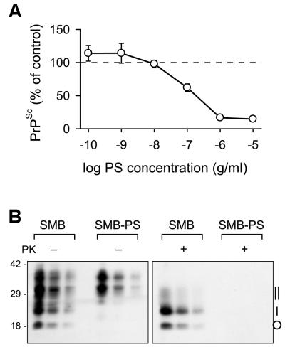
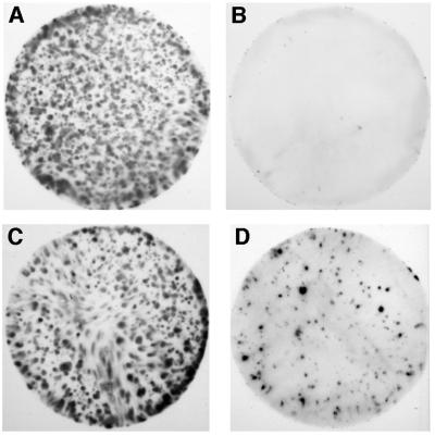
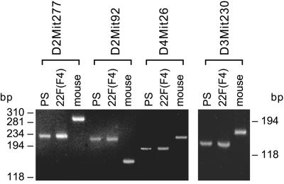
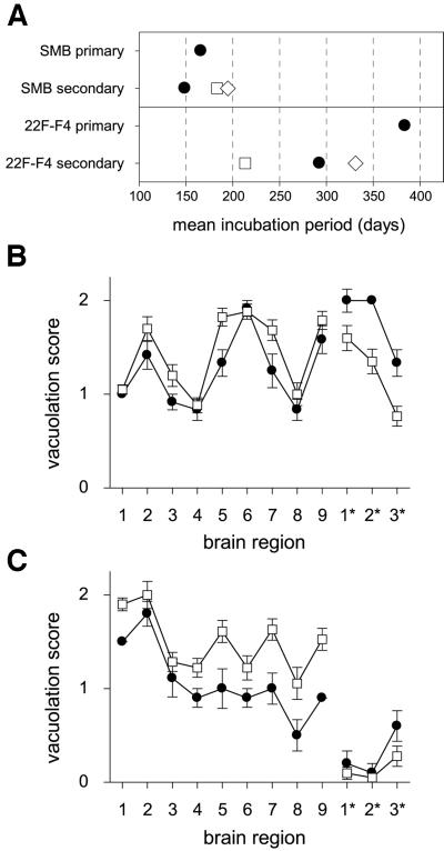
Bovine spongiform encephalopathy (BSE) and its human equivalent, variant Creutzfeldt-Jakob disease (vCJD), are caused by the same strain of infectious agent, which is similar to, but distinct from, >20 strains of their sheep scrapie homologue. A better understanding of the molecular strain determinants could be obtained from cells in monoculture than from whole animal studies where different cell targeting is commonly a strain-related feature. Although a few cell types can be infected with different strains, the phenotypes of the emergent strains have not been studied. We have cured the scrapie-infected, clonal SMB cell line with pentosan sulfate, stably re-infected it with a different strain of scrapie and shown that biological properties and prion protein profiles characteristic of each original strain are propagated faithfully in this single non-neuronal cell type. These findings attest to the fact that scrapie strain determinants are stable and host-independent in isolated cells.


Figures

None
Fig. 1. PrPSc expression in SMB cells was cured by pentosan sulfate. (A) The inhibition by PS of PrPSc accumulation in SMB cells is dose dependent. Cultures of SMB cells were grown for 7 days in the continuous presence of the indicated concentrations of PS, after which time PrPSc levels were measured by quantitative dot-blotting. The points are the means (±SEM) from four experiments in which each data point was triplicated. (B) Western blot analysis of the PrP in SMB and the pentosan sulfate-treated SMB-PS cells. Proteins in post-nuclear cell extracts prepared either with (+) or without (–) proteinase K (PK) treatment, were precipitated with methanol/chloroform prior to electrophoresis. Successive lanes in each triplet of lanes represents a 5-fold dilution series of the previous sample. Molecular weight standards (kDa) are indicated on the left and the position of PK-treated PrPSc isoforms on the right: non-glycosylated (circle), monoglycosylated (single vertical bar) and diglycosylated (double vertical bar).
None
Fig. 2. Scrapie brain but not normal brain stimulates PrPSc synthesis after in vitro challenge. SMB-PS cells were exposed to a homogenate of either normal mouse brain (A and B) or 22F-infected brain (C and D); the donor mice were of the VM(Sincs7) strain. Filters from identical cultures were subjected to development with anti-PrP antibody (IA8) either without proteinase K (PK) treatment, giving the total clone density (A and C), or after treatment with 100 µg/ml PK (B and D), showing only those clones containing PrPSc.
None
Fig. 3. The same lineage of SMB-PS and 22F-infected cells was determined by microsatellite analysis. PCR-microsatellite analysis using the indicated primers was performed on DNA from SMB-PS cells (PS), 22F-challenged SMB-PS cells, clone F4 [22F (F4)] and the VM(Sincs7) mouse (mouse), the 22F-infected donor. Size calibration in base pairs (bp) is shown to left and right.
None
Fig. 4. Strain typing of cell-derived scrapie revealed distinctive characteristics. (A) Comparative ranking of the scrapie incubation periods in mice at the primary and secondary transmissions. Primary transmissions were established by injecting 7.5 × 10⁴ cell equivalents intracerebrally into mice, and secondary transmissions were made by injecting 0.03 ml of a 10–2 homogenate of the brain taken from a single primary transmission mouse once it had reached the clinical stage of the disease. Strains of mice used were C57BL (Sincs7; filled circles), VM (Sincp7; open squares) and their F1 cross (Sincs7p7; open diamonds). Each point is the mean of 12 mice (primary transmissions) or 20–24 mice (secondary transmissions); the error bars fall within the boundaries of the symbols and are therefore not seen. (B and C) Lesion profiles generated by the primary (filled circles) and secondary transmission (open squares) of cell-borne scrapie; from SMB cells (B) or from SMB[22F]F4 cells (C). In both (B) and (C), each point is the mean score at the specified brain region of between 12 and 22 animals, the bars are 1 SEM. Grey matter regions scored: 1, dorsal medulla; 2, cerebellar cortex; 3, superior colliculus; 4, hypothalamus; 5, medial thalamus; 6, hippocampus; 7, septum; 8, thalamic cortex; 9, forebrain cortex. White matter regions scored: 1*, cerebellar white matter; 2*, mesencephalic tegmentum; 3*, pyramidal tract.
None
Fig. 5. PrPSc isoform analysis of infected cells revealed distinct differences both between strains and between cells and brains. (A) Western blotting was used to compare PK-treated PrPSc derived from mouse brain with that from SMB and SMB[22F]F4 cells. The lanes were loaded as follows: lane 1, 139A reference mouse brain sample; lane 2, SMB cells, ∼8 × 10⁵ cell equivalents; lane 3, mouse brain from primary transmission of SMB cells; lane 4, mouse brain from secondary transmission of SMB cells; lane 5, 22F reference mouse brain sample; lane 6, SMB[22F]F4 cells, ∼3 × 10⁵ cell equivalents; lane 7, mouse brain from primary transmission of SMB[22F]F4 cells; and lane 8, mouse brain from secondary transmission of SMB[22F]F4 cells. Each brain sample was ∼40 µg wet weight of tissue. Molecular weight standards (kDa) are marked on the left. (B) Densitometric analysis constructed from a number of western-blotted samples developed with the IA8 antiserum. Grouped bars depicting the diglycosyl (filled bar), monoglycosyl (hatched bar) and unglycosylated (open bar) PrPSc isoforms are numbered the same as in (A). The columns are the mean of between six and 12 brains (samples 1, 3–5, 7 and 8) or from 11 or 14 cell samples (samples 2 and 6, respectively) collected intermittently over a 50 passage series; the bars represent 1 SD.

Similar articles

Cited by

References

    1. Aguzzi A. and Weissmann,C. (1997) Prion research: the next frontiers. Nature, 389, 795–798. - PubMed
    1. Bessen R.A. and Marsh,R.F. (1994) Distinct PrP properties suggest the molecular basis of strain variation in transmissible mink encephalopathy. J. Virol., 68, 7859–7868. - PMC - PubMed
    1. Bessen R.A., Kocisko,D.A., Raymond,G.J., Nandan,S., Lansbury,P.T. and Caughey,B. (1995) Non-genetic propagation of strain-specific properties of scrapie prion protein. Nature, 375, 698–700. - PubMed
    1. Bosque P.J. and Prusiner,S.B. (2000) Cultured cell sublines highly susceptible to prion infection. J. Virol., 74, 4377–4386. - PMC - PubMed
    1. Brimacombe D.B., Bennett,A.D., Wusteman,F.S., Gill,A.C., Dann,J.C. and Bostock,C.J. (1999) Characterization and polyanion-binding properties of purified recombinant protein. Biochem. J., 342, 605–613. - PMC - PubMed

Publication types