. 2001 Jul 2;20(13):3370-9.
doi: 10.1093/emboj/20.13.3370.

Analysis of heterodimer formation by Xklp3A/B, a newly cloned kinesin-II from Xenopus laevis

Affiliations

Analysis of heterodimer formation by Xklp3A/B, a newly cloned kinesin-II from Xenopus laevis

V De Marco et al. EMBO J. .

Abstract

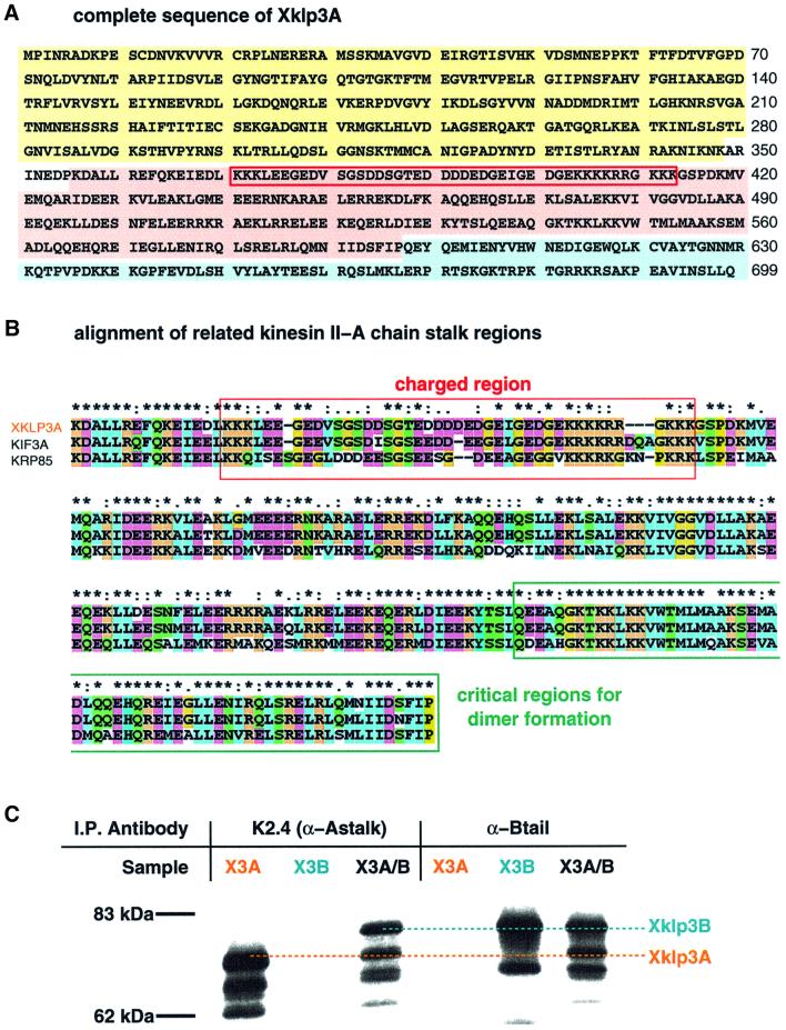
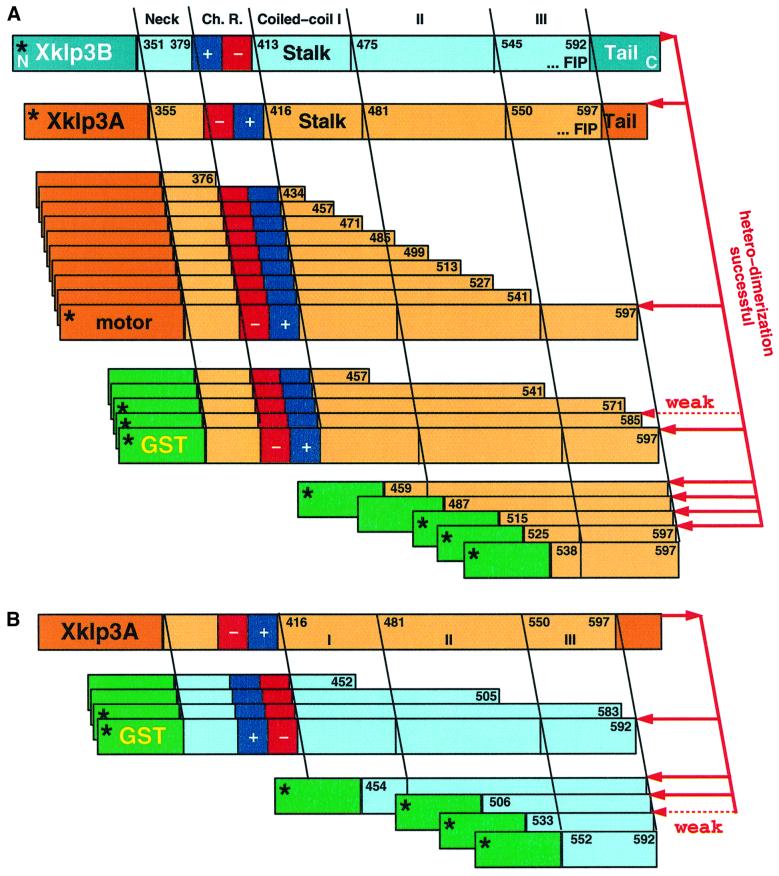
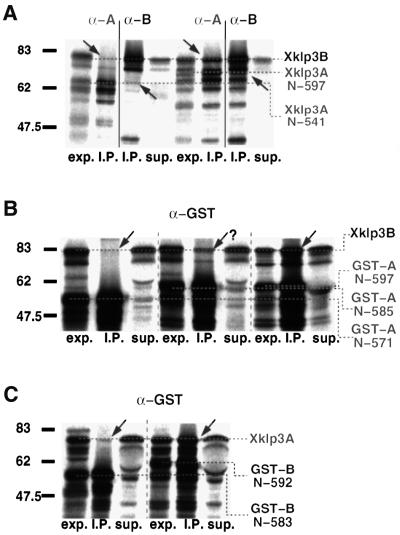
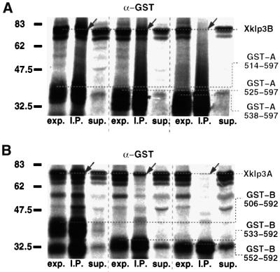
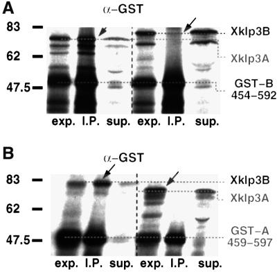
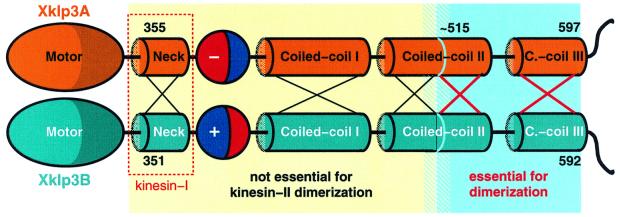
kinesin-II motor proteins are composed of two different kinesin-like motor proteins and one cargo binding subunit. Here we report the cloning of a new member of the kinesin-II superfamily, Xklp3A from Xenopus laevis, which forms a heterodimeric complex with Xklp3B. The heterodimer formation properties between Xklp3A and B have been tested in vitro using reticulocyte lysate expression and immunoprecipitation. To this end we produced a series of Xklp3A and B constructs of varying length and tested their propensity for heterodimer formation. We could demonstrate that, in contrast to conventional kinesin, the critical domains for heterodimer formation in Xklp3A/B are located at the C-terminal end of the stalk. Neither the neck nor the highly charged stretches after the neck region, which are typical of kinesins-II, are required for heterodimer formation, nor do they prevent homodimer formation. Dimerization is controlled by a cooperative mechanism between the C-terminal coiled-coil segments. Classical trigger sites were not identified. The critical regions for dimerization exhibit a very high degree of sequence conservation among equivalent members of the kinesin-II family.


Figures

None
Fig. 1. Xklp3A belongs to the kinesin II family and forms a complex with Xklp3B. The predicted protein sequence of Xklp3A is shown in (A). Domains are marked as follows: motor domain, yellow; stalk region, pink; tail domain, blue. A red frame marks the stretch of highly charged amino acids that separates the neck from the coiled-coil stalk. (B) The predicted sequence for the stalk region in Xklp3A is aligned with MmKIF3A and SpKRP85: identical residues are marked with an asterisk (∗), conserved residues with double dots (:), semi-conserved residues with a single dot (.). The colour code distinguishes between non-polar amino acids (blue), polar (green) and charged residues (orange for positively charged, purple for negatively charged). The alignment was performed using ClustalX. Interestingly the critical region for dimerization (C-terminal part of the stalk) is highly conserved among related members of the kinesin II family. (C) Immunoprecipitation of Xklp3A/B heterodimer: an α-Astalk antibody (K2.4: raised against the stalk of SpKRP85) precipitates Xklp3A but not Xklp3B unless they are co-expressed and a complex between the two kinesin-like proteins is formed. Consequently an α-Xklp3B antibody (α-Btail) immunoprecipitates Xklp3A only when expressed with Xklp3B.
None
Fig. 2. Xklp3A and Xklp3B show a tripartite organization of the stalk domain. Heptad-repeat assignment for Xklp3A and Xklp3B stalks, as predicted by Multicoil and visual inspections, reveals three coiled-coil subdomains, connected through stutters (Brown et al., 1996). The neck region is separated from the coiled-coil stalk by the charged region. The neck and in particular the charged regions are not predicted to fold in a stable coiled coil. Arrows mark repulsive e–g ionic interactions in Xklp3A coiled-coil region II, which could prevent homodimerization.
None
Fig. 3. Schematic overview of the constructs, which were probed for heterodimer formation. (A) Heterodimer formation of full-length Xklp3B chain was tested against Xklp3A constructs with various stalk fragments. Xklp3A/B heterodimer formation occurs only when the C-terminal part of Xklp3A stalk is present. N-terminal truncations allow complete omission of the neck, the charged regions (Ch. R.), coiled-coil region I and most of coiled-coil region II. In some experiments the motor head was exchanged with a GST domain, which allowed for a more effective immunoprecipitation. This did not change the outcome of the precipitation experiments and revealed identical results (see Figures 4–6). (B) The reversed experimental set-up of (A). Confirming the findings made with Xklp3A segments, the C-terminal part of Xklp3B stalk is equally essential for heterodimer formation. The asterisks mark constructs of which immunoprecipitation results are shown in Figures 4–6.
None
Fig. 4. Heterodimer formation is ‘triggered’ by the C-terminal part of the stalk. (A) The removal of even a short part of the C-terminal end of the Xklp3A stalk inhibits heterodimer formation. Xklp3B was co-expressed with Xklp3A:N-541 (left lanes) or Xklp3A:N-597 (right lanes). Complex formation was monitored by immunoprecipitation using both α-SpKRP85 (α-A) and α-Xklp3B (α-B) antibodies. The construct with full-length stalk is the only one precipitated through heterodimer formation. (B) Even short truncations of the Xklp3A coiled-coil region III from the C-terminal end inhibit coiled-coil formation. Xklp3B was co-expressed with GST-A:N-571, GST-A:N-585 and GST-A:N-597. Only GST-A:N-597 is fully capable of precipitating Xklp3B. Complex formation was tested by immunoprecipitation with an α-GST antibody. (C) In analogy to the experiments shown in (B), truncation of the Xklp3B coiled-coil region III destabilizes dimer formation in a very similar way. Xklp3A was co-expressed with GST-B:N-583 or GST-B:N-592 and precipitated with α-GST. GST-B:N-583 is already too short to form a heterodimeric complex. Lanes are marked as follows: exp., lysate before immunoprecipitation; I.P., immunoprecipitate; sup., supernatant after precipitation. Arrows mark the position of products coprecipitated via heterodimer formation. Compare also with Figure 3.
None
Fig. 5. Heterodimer formation is controlled by a cooperative interaction between coiled-coil regions II and III. (A) Xklp3B was co-expressed with GST-A:514–597 (leaves five complete C-terminal heptads of coiled-coil region II), GST-A:525–597 (four heptads left) or with GST-A:538–597 (two heptads left). GST-A:525–597 shows a lower efficiency in complex formation, and GST-A:538–597 is incapable of forming a heterodimer. As in Figure 4B and C heterodimer formation was monitored by immunoprecipitation with an α-GST antibody. (B) Reversing the conditions from (A), Xklp3A was co-expressed with GST-B:506–592 (six heptads left), GST-B:533–592 (two heptads left) or GST-B:552–592 (complete coiled-coil region III). In this case slightly shorter stretches are still showing some heterodimer formation. GST-B:506–592 is fully capable, GST-B:533–592 is much less capable and GST-B:552–592 is incapable of heterodimer formation. Lanes are marked as follows: exp., lysate before immunoprecipitation; I.P., immunoprecipitate; sup., supernatant after precipitation. Arrows mark the position of products coprecipitated via heterodimer formation.
None
Fig. 6. The removal of Xklp3A and Xklp3B charged regions does not affect heterodimer formation and does not promote homodimerization of the two subunits. (A) GST-B:454–592, lacking the N-terminal regions between the beginning of the neck and the end of the charged regions, was co-expressed either with Xklp3A or Xklp3B. As in Figures 3 and 4 complex formation was checked by immunoprecipitation with α-GST. Complex formation occurs only between GST-B:454–592 and Xklp3A (left lanes) but not between GST-B:454–592 and Xklp3B (right). (B) Vice versa, GST-A:459–597 was co-expressed with either Xklp3A or Xklp3B. As in (A) α-GST precipitates only a complex that is formed between GST-A:459–597 and Xklp3B (left) but not between GST-A:459–597 and Xklp3A (right). Lanes are marked as follows: exp., lysate before immunoprecipitation; I.P., immunoprecipitate; sup., supernatant after precipitation. Arrows mark the position of products coprecipitated via heterodimer formation.
None
Fig. 7. Xklp3A/B heterodimer formation appears to be based on a cooperative mechanism between the C-terminal part of coiled-coil region II and the entire coiled-coil region III. Coiled-coil region II needs to maintain a minimum length of about two to three heptad repeats otherwise heterodimer formation is inhibited. Four remaining heptads restore full activity. Here we illustrate the key difference between dimerization of kinesin-II and of conventional kinesin. In contrast to the situation in conventional kinesin, the neck regions (red box) are not responsible for dimerization. Xklp3A/B heterodimerization is driven by the C-terminal part of the stalk. The sequences at the N- or C-terminal end of the blue region do not exhibit any coiled-coil trigger sequences. The absence of trigger sites and the involvement of two separated coiled-coil regions strongly suggest that dimerization is mediated by a cooperative mechanism that raises the relatively low individual coiled-coil-forming propensities of regions II and III to a physiologically significant level.

Similar articles

Cited by

References

    1. Arndt K.M., Pelletier,J.N., Muller,K.M., Alber,T., Michnick,S.W. and Pluckthun,A. (2000) A heterodimeric coiled-coil peptide pair selected in vivo from a designed library-versus-library ensemble. J. Mol. Biol., 295, 627–639. - PubMed
    1. Brady S.T. (1985) A novel brain ATPase with properties expected for the fast axonal transport motor. Nature, 317, 73–75. - PubMed
    1. Brown J.H., Cohen,C. and Parry,D.A. (1996) Heptad breaks in α-helical coiled coils: stutters and stammers. Proteins, 26, 134–145. - PubMed
    1. Brown J.M., Marsala,C., Kosoy,R. and Gaertig,J. (1999) Kinesin-II is preferentially targeted to assembling cilia and is required for ciliogenesis and normal cytokinesis in Tetrahymena. Mol. Biol. Cell, 10, 3081–3096. - PMC - PubMed
    1. Burkhard P., Kammerer,R.A., Steinmetz,M.O., Bourenkov,G.P. and Aebi,U. (2000a) The coiled-coil trigger site of the rod domain of cortexillin I unveils a distinct network of interhelical and intrahelical salt bridges. Struct. Fold. Des., 8, 223–230. - PubMed

MeSH terms