. 2001 Jul 2;20(13):3414-26.
doi: 10.1093/emboj/20.13.3414.

PTP-PEST, a scaffold protein tyrosine phosphatase, negatively regulates lymphocyte activation by targeting a unique set of substrates

Affiliations

PTP-PEST, a scaffold protein tyrosine phosphatase, negatively regulates lymphocyte activation by targeting a unique set of substrates

D Davidson et al. EMBO J. .

Abstract

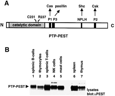
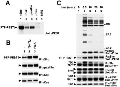
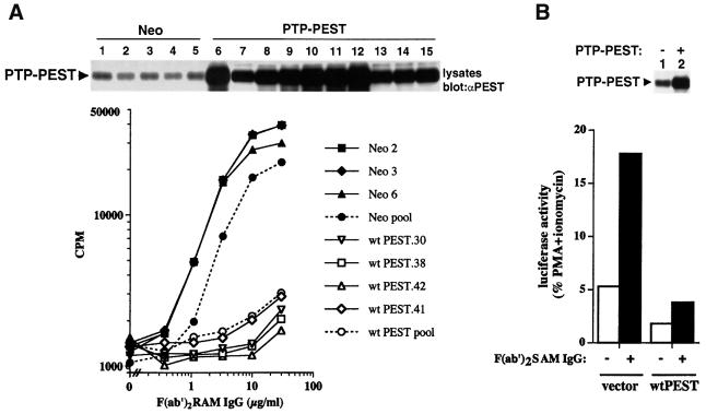
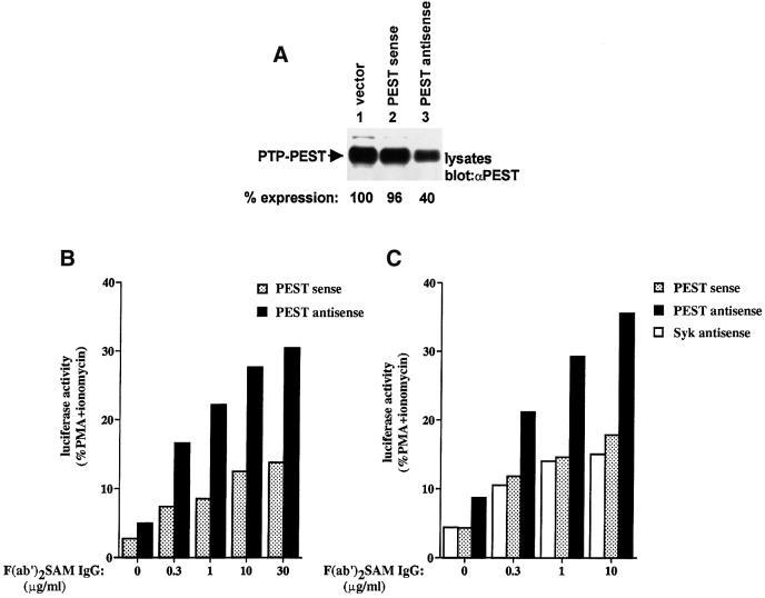
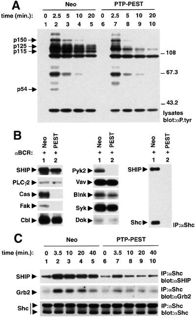
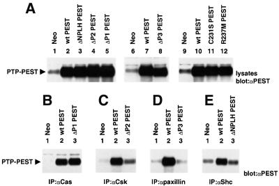
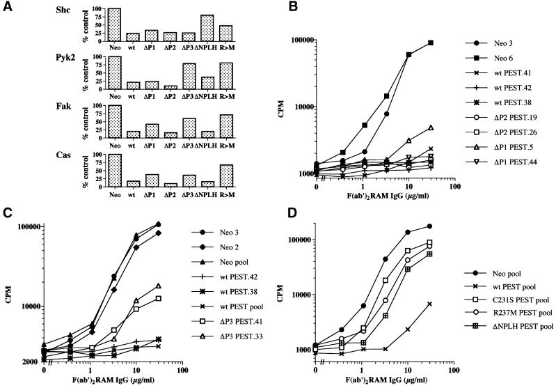
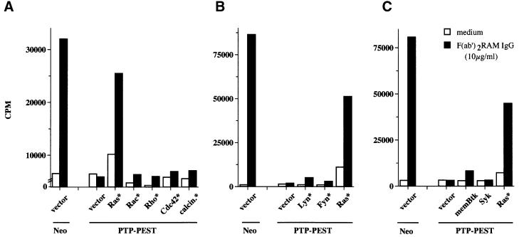
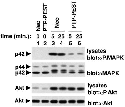
There is increasing interest in elucidating the mechanisms involved in the negative regulation of lymphocyte activation. Herein, we show that the cytosolic protein tyrosine phosphatase PTP-PEST is expressed abundantly in a wide variety of haemopoietic cell types, including B cells and T cells. In a model B-cell line, PTP-PEST was found to be constitutively associated with several signalling molecules, including Shc, paxillin, Csk and Cas. The interaction between Shc and PTP-PEST was augmented further by antigen receptor stimulation. Overexpression studies, antisense experiments and structure-function analyses provided evidence that PTP-PEST is an efficient negative regulator of lymphocyte activation. This function correlated with the ability of PTP-PEST to induce dephosphorylation of Shc, Pyk2, Fak and Cas, and inactivate the Ras pathway. Taken together, these data suggest that PTP-PEST is a novel and unique component of the inhibitory signalling machinery in lymphocytes.


Figures

None
Fig. 1. Structure and expression pattern of PTP-PEST in haemopoietic cells. (A) The primary structure of PTP-PEST is depicted graphically. Two residues in the catalytic domain (Cys231 and Arg237), that are critical for phosphatase activity, are highlighted. The positions of the P1, P2, P3 and NPLH sequences in the C-terminal non-catalytic domain are also shown. (B) Expression pattern of PTP-PEST in mouse haemopoietic cells. The accumulation of PTP-PEST in various purified populations of mouse haemopoietic cells was examined by immunoblotting of equivalent amounts of cell lysates with a rabbit anti-PTP-PEST (PEST) serum. The identity of the higher molecular weight polypeptides found to react with the anti-PTP-PEST sera in this immunoblot is not known. The presence of comparable quantities of cellular proteins in each lane was verified by staining of the immunoblot membrane with Coomassie blue (data not shown). The position of PTP-PEST is indicated on the left. Exposure: 25 s (ECL).
None
Fig. 2. Association of PTP-PEST with signalling molecules in mouse A20 B cells. IgG+ A20 B cells were lysed in NP-40-containing buffer, and the ability of PTP-PEST to associate with other polypeptides was determined by immunoblotting of the indicated immunoprecipitates with an anti-PTP-PEST (PEST) serum. The position of PTP-PEST is highlighted by an arrow on the left. (A) Association of PTP-PEST with signalling molecules in unstimulated A20 cells. NRS: normal rabbit serum. Exposure: 60 s (ECL). (B) Effects of BCR stimulation or PMA treatment on the association of PTP-PEST with signalling molecules. A20 cells were either left unstimulated or stimulated for 10 min with F(ab′)2 fragments of SAM IgG antibodies (20 µg/ml) or PMA (100 ng/ml), prior to cell lysis and immunoprecipitation. Exposures: first panel, 3 h; second, third and fourth panels, 10 s (ECL). (C) Time course of BCR stimulation. Cells were stimulated for the indicated times with F(ab′)2 fragments of SAM IgG antibodies. The accumulation of phosphotyrosine-containing proteins was monitored by immunoblotting of total cell lysates with anti-phosphotyrosine (P.tyr) antibodies (first panel), whereas the presence of Shc–PTP-PEST complexes was detected by immunoblotting of Shc immunoprecipitates with an anti-PTP-PEST serum (second panel). Tyrosine phosphorylation of total Shc (third and fourth panels) and PTP-PEST-bound Shc (fifth and sixth panels) was analysed in parallel. Note that whereas both the 52 and 46 kDa Shc isoforms were recovered in Shc immunoprecipitates (fourth panel), only the 52 kDa variant was associated with PTP-PEST (sixth panel). This presumably is because the 46 kDa Shc protein lacks part of the PTB domain, which mediates the binding to PTP-PEST. The migration of pre-stained molecular mass markers is indicated on the right. Exposures: first panel, 14 h; second panel, 4 h; third panel, 15 h; fourth panel, 2.5 h; fifth panel, 15 h; sixth panel, 2 h.
None
Fig. 3. Effect of PTP-PEST on antigen receptor-induced activation. (A) Stable overexpression of PTP-PEST in A20 B cells. Stable transfectants overexpressing wild-type PTP-PEST (wt PEST) or expressing the neomycin resistance marker alone (Neo) were stimulated with the indicated concentrations of F(ab′)2 fragments of RAM IgG antibodies (abscissa), as detailed in Materials and methods. The accumulation of IL-2 in the supernatant was assessed by measuring tritiated thymidine incorporation in the IL-2-dependent indicator cell line HT-2 (ordinate). All assays were done in triplicate and repeated at least 10 times. The quantities of PTP-PEST contained in the various cell lines used in this report were determined by immunoblotting of total cell lysates with anti-PTP-PEST antibodies (top panel). Lane 1, Neo.1; lane 2, Neo.2; lane 3, Neo.3; lane 4, Neo.5; lane 5, Neo.6; lane 6, wtPEST.70 (18); lane 7, wtPEST.35 (4); lane 8, wtPEST.30 (9); lane 9, wtPEST.38 (11); lane 10, wtPEST.42 (15); lane 11, wtPEST.64 (16); lane 12, wtPEST.70 (18); lane 13, wtPEST.31 (4.5); lane 14, wtPEST.41 (5); lane 15, wtPEST.71 (6) (fold PTP-PEST overexpression is shown in parentheses). The position of PTP-PEST is indicated on the left. Exposure: 6 h. (B) Transient overexpression of PTP-PEST in A20 B cells. A20 cells were transiently transfected with the vector pXM139 alone or bearing a wild-type mouse ptp-pest cDNA, in the presence of an IL-2 promoter–luciferase reporter construct. Cells were then stimulated with F(ab′)2 fragments of SAM IgG antibodies for 6 h, and luciferase activity was measured in a luminometer as detailed in Materials and methods. Results are represented as the percentage of maximal stimulation obtained with PMA plus ionomycin. Expression levels of PTP-PEST are shown at the top. Exposure: 3 h.
None
Fig. 4. Expression of ptp-pest antisense in A20 B cells. (A) Effect of antisense ptp-pest expression on the abundance of PTP-PEST in A20 cells. A20 cells were transiently transfected with 40 µg of empty pXM139 or pXM139 bearing the first 321 nucleotides of the coding sequence of a mouse ptp-pest cDNA, in either the antisense or the sense orientation. After 48 h, equivalent numbers of viable cells were lysed and the abundance of PTP-PEST was examined by immunoblotting of total cell lysates with anti-PTP-PEST antibodies. The migration of PTP-PEST is indicated on the left. Exposure: 7 h. (B) Influence of antisense ptp-pest expression on BCR signalling. A20 cells were transfected as detailed in (A), in the presence of the IL-2 promoter–luciferase reporter construct. After 66 h, they were stimulated with F(ab′)2 fragments of SAM IgG antibodies for 6 h, and luciferase activity was measured as explained in Materials and methods. Data are shown as the percentage of maximal stimulation achieved with PMA plus ionomycin. (C) Impact of antisense syk expression on BCR signalling. As in (B), except that an antisense syk construct was also used. In keeping with the positive regulatory role of Syk in BCR signalling, a small decrease in activation of the reporter plasmid was observed in BCR-stimulated A20 cells expressing the antisense syk plasmid.
None
Fig. 5. Impact of PTP-PEST overexpression on BCR-induced protein tyrosine phosphorylation. (A) Overall protein tyrosine phosphorylation. Representative cell lines expressing the neomycin marker alone (Neo; lanes 1–5) or in combination with wild-type PTP-PEST (lanes 6–10) were stimulated for the indicated times with F(ab′)2 fragments of SAM IgG antibodies. Changes in intracellular protein tyrosine phosphorylation were assessed by immunoblotting of total cell lysates with anti-phosphotyrosine (P.tyr) antibodies. The migrations of pre-stained molecular mass markers are shown on the right, while those of p150, p125, p115 and p54 are indicated on the left. Exposure: 15 h. (B) Tyrosine phosphorylation of specific substrates. Cells expressing the neomycin marker alone (Neo; lane 1) or in combination with wild-type PTP-PEST (PEST; lane 2) were activated for 2.5 min with F(ab′)2 fragments of SAM IgG antibodies. Specific substrates were then immunoprecipitated from cell lysates using the appropriate antibodies, and their phosphotyrosine content was determined by anti-phospho tyrosine (P.tyr) immunoblotting. Equivalent amounts of the various substrates were immunoprecipitated in control cells and in cells overexpressing PTP-PEST (data not shown). The positions of the various polypeptides are indicated on the left. Exposures: SHIP, 9 h; PLCγ2, 30 h; Cas, 21 h; Fak, 30 h; Cbl, 9 h; Pyk2, 9 h; Vav, 9 h; Blnk, 30 h; Syk, 12 h; Dok, 12 h; and Shc, 12 h. (C) Effect of PTP-PEST overexpression on association of Shc with SHIP and Grb2. Cells were activated for the specified times. The extent of association of Shc with SHIP and Grb2 was established by immunoblotting of anti-Shc immunoprecipitates with anti-SHIP (first panel) or anti-Grb2 (second panel) antibodies. The abundance of Shc was also verified by anti-Shc immunoblotting of Shc immunoprecipitates (third panel). The migrations of SHIP, Grb2 and Shc are noted on the left. Exposures: first panel, 13 h; second panel, 39 h; third panel, 2.5 h.
None
Fig. 6. Structural requirements for inhibition of antigen receptor signalling by PTP-PEST. (A) Expression of PTP-PEST mutants in A20 cells. The expression levels of the various PTP-PEST mutants were determined by immunoblotting of total cell lysates from representative cell lines with anti-PTP-PEST (PEST) antibodies. The migration of PTP-PEST is shown on the left. Exposures: lanes 1–5, 3 h; lanes 6–8, 3 h; lanes 9–12, 6 h. (BE) Impact of mutations on the ability of PTP-PEST to associate with signalling molecules. The capacity of the PTP-PEST mutants to bind other polypeptides was assessed by immunoblotting of the appropriate immunoprecipitates with anti-PTP-PEST antibodies. The associations of the PTP-PEST mutants with other binding proteins not shown here were not affected (data not shown). The migration of PTP-PEST is indicated on the left. Exposures: (B) 8 h; (C) 3 h; (D) 6 h; and (E) 4 h.
None
Fig. 7. Influence of PTP-PEST mutants on BCR-induced protein tyrosine phosphorylation and IL-2 production. (A) BCR-induced protein tyrosine phosphorylation. The extent of BCR-induced tyrosine phosphorylation of individual substrates in A20 derivatives expressing the various PTP-PEST polypeptides was compared with that observed in Neo cells. Data from several independent experiments were quantitated with a phosphoimager, and are represented graphically. (BD) BCR-triggered lymphokine production. Cells were tested as detailed in Figure 3A. In some cases, pools of transfectants were assayed. All assays were done in triplicate and repeated a minimum of three times.
None
Fig. 8. Selective rescue of PTP-PEST-mediated inhibition by activated Ras. (AC) Rescue of the PTP-PEST-induced block by activated Ras. Pools of A20 transfectants stably overexpressing wild-type PTP-PEST were transiently transfected with the indicated constructs. Cells subsequently were activated with F(ab′)2 fragments of RAM IgG antibodies, and IL-2 release in the supernatant was measured using a bioassay. Cells expressing the neomycin phosphotransferase alone (Neo) were transfected with empty vector alone, and were used as control. All assays were done in triplicate. Expression of all constructs was confirmed by immunoblotting of total cell lysates with the appropriate antibodies (data not shown).
None
Fig. 9. Impact of PTP-PEST overexpression on MAPK activation. Cells expressing the neomycin resistance marker alone (Neo) or in combination with wild-type PTP-PEST were stimulated for the indicated times with F(ab′)2 fragments of SAM IgG antibodies (0.37 µg/ml). Activation of MAPK (Erk) was assayed by immuno blotting of total cell lysates with antibodies specific for the activated form of MAPK (first panel). Levels of expression of MAPK were confirmed by immunoblotting of parallel lysates with anti-MAPK antibodies (second panel). Activation of Akt was studied as control, using an antibody specific for activated Akt (third panel). Levels of Akt were determined by immunoblotting with an anti-Akt antibody (fourth panel). The positions of p42MAPK (Erk-2), p44MAPK (Erk-1) and Akt are indicated on the left. Exposures: first panel, 4 h; second panel, 4 h; third panel, 48 h; fourth panel, 3 min (ECL).

Similar articles

Cited by

References

    1. Angers-Loustau A., Coté,J.F., Charest,A., Dowbenko,D., Spencer,S., Lasky,L.A. and Tremblay,M.L. (1999) Protein tyrosine phosphatase-PEST regulates focal adhesion disassembly, migration and cytokinesis in fibroblasts. J. Cell Biol., 144, 1019–1031. - PMC - PubMed
    1. Benschop R.J. and Cambier,J.C. (1999) B cell development: signal transduction by antigen receptors and their surrogates. Curr. Opin. Immunol., 11, 143–151. - PubMed
    1. Bolland S., Pearse,R.N., Kurosaki,T. and Ravetch,J.V. (1998) SHIP modulates immune receptor responses by regulating membrane association of Btk. Immunity, 8, 509–516. - PubMed
    1. Campbell K.S. (1999) Signal transduction from the B cell antigen-receptor. Curr. Opin. Immunol., 11, 256–264. - PubMed
    1. Cao M.Y., Davidson,D., Yu,J., Latour,S. and Veillette,A. (1999) Clnk, a novel SLP-76-related adaptor molecule expressed in cytokine-stimulated hemopoietic cells. J. Exp. Med., 190, 1527–1534. - PMC - PubMed

Publication types

MeSH terms

Substances