. 2001 Jul 2;20(13):3437-46.
doi: 10.1093/emboj/20.13.3437.

A direct interaction between JNK1 and CrkII is critical for Rac1-induced JNK activation

Affiliations

A direct interaction between JNK1 and CrkII is critical for Rac1-induced JNK activation

S E Girardin et al. EMBO J. .

Abstract

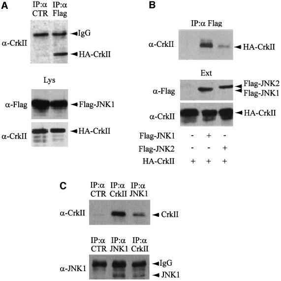
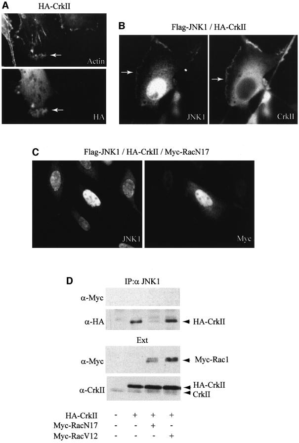
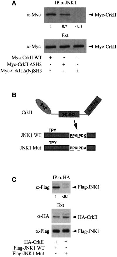
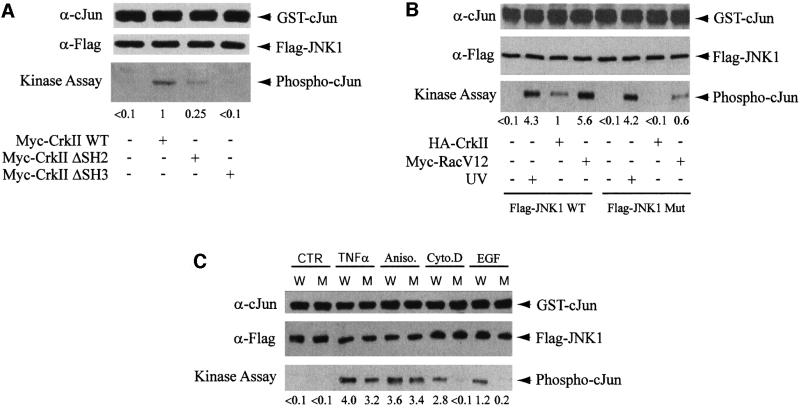
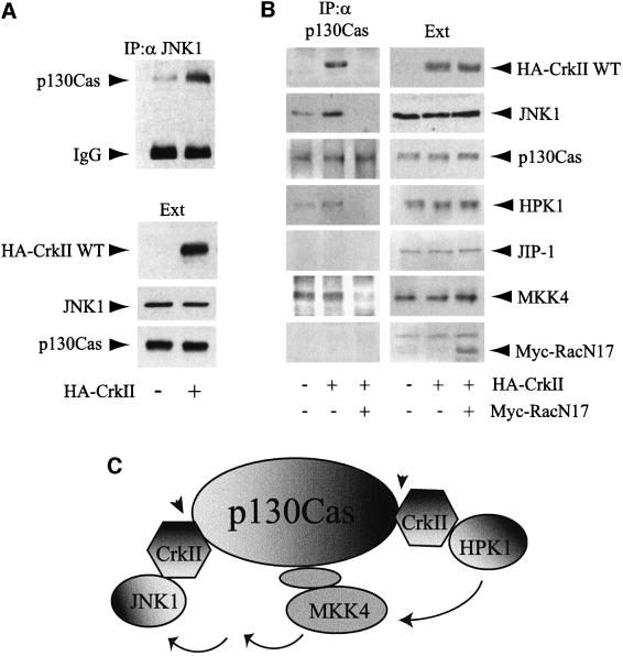
CrkII, a cellular homolog of v-crk, belongs to a family of adaptor proteins that play a central role in signal transduction cascades. We demonstrate that CrkII interacts directly with c-Jun N-terminal kinase 1 (JNK1). A proline-rich sequence of JNK1 is critical for the interaction of the kinase with the N-terminal Src homology 3 (SH3) domain of CrkII. JNK1 is localized with CrkII in membrane ruffles of Crk-overexpressing cells in a Rac1-dependent manner. A JNK1 mutant (K340A) that fails to interact with CrkII is defective in Rac/epidermal growth factor-induced activation, but remains responsive to UVC irradiation. Furthermore, CrkII recruits JNK1 to a p130Cas multiprotein complex where it may be activated through a hematopoietic progenitor kinase 1- and mitogen-activated protein kinase kinase 4-dependent pathway. Together, the results presented here argue for a new mechanism of regulation of the JNK pathway through the CrkII-p130Cas adaptor complex.


Figures

None
Fig. 1. CrkII interacts with JNK1. (AIn vitro interaction. Flag-JNK1 and HA-CrkII were prepared by in vitro translation using wheat germ extracts, mixed, and separated into two aliquots (Lys). A fraction of each aliquot was fractionated on polyacrylamide gel, transferred to a membrane and blotted with either anti-Flag or anti-CrkII antibodies (lower panel). The rest of the two aliquots were immunoprecipitated with an unrelated mouse monoclonal antibody (CTR, left lane) or mouse monoclonal anti-Flag antibody (right lane) and probed with anti-CrkII antibody (upper panel). (BIn vivo interaction. The plasmids encoding HA-CrkII, Flag-JNK1 or Flag-JNK2 were cotransfected into HeLa cells as indicated. The yield of transfected proteins in the extracts (Ext) was determined after immunoblotting with either anti-Flag or anti-CrkII antibodies (lower panel). Interaction was determined by immunoprecipitation using anti-Flag mouse monoclonal antibody. The presence of CrkII in the Flag immunoprecipitates was determined using an anti-CrkII antibody (upper panel). (C) Interaction of the endogenous proteins. The interaction between CrkII and JNK1 was directly addressed with the endogenous proteins after JNK1 or CrkII immunoprecipitation followed by western blotting carried out on HeLa cellular extracts. Immunoprecipitations using an unrelated antibody (CTR) are also presented as negative control.
None
Fig. 2. CrkII interaction with JNK1, and CrkII-dependent JNK1 localization to ruffles are regulated by Rac1 activity. (A) Overexpression of CrkII induces membrane ruffling. HeLa cells were visualized by indirect immunofluorescence after transfection with HA-CrkII expression vector. Cells were stained using a mouse monoclonal anti-HA. Rhodamine-conjugated phalloidin was used to visualize filamentous actin. Membrane ruffling induced by CrkII is indicated by an arrow. (B) JNK1 localization to ruffles. HeLa cells were visualized by indirect immunofluorescence after cotransfection with Flag-JNK1 and HA-CrkII expression vectors. Cells were stained using a mouse monoclonal anti-CrkII or a polyclonal rabbit anti-JNK1 antibody as indicated. JNK1 localization to ruffles is indicated by an arrow. (C) Effect of Rac1 on JNK1 localization to ruffles. HeLa cells were visualized by indirect immunofluorescence after cotransfection with Flag-JNK1, HA-Crk and Myc-RacN17 expression vectors. Cells were stained using a rabbit polyclonal anti-JNK1 antibody (left panel) or a mouse monoclonal anti-Myc antibody (right panel). Membrane ruffles were absent in all the cells transfected with Myc-RacN17 expression vector. (D) Effect of Rac1 on the CrkII–JNK1 interaction. HeLa cells were transfected with HA-CrkII plus either Myc-RacN17 or Myc-RacV12 expression vectors as indicated. Interaction between JNK1 and transfected HA-CrkII was determined by HA immunoblot on JNK1 immunoprecipitates (top). Myc-Rac1 proteins were not found in the JNK1 immunoprecipitates. Control immunoblots on total protein extracts (Ext) using anti-Myc or anti-CrkII antibodies are presented.
None
Fig. 3. JNK1 interacts with CrkII through CrkII N-terminal SH3 domain. (A) HeLa cells were transfected with Myc-CrkII, Myc-CrkΔSH2 or Myc-CrkΔ(N)SH3 expression vectors as indicated. Interaction between JNK1 and the different forms of CrkII was determined by Myc immunoblot on JNK1 immunoprecipitates. The numbers below the blot give the yield. (B) Schematic diagram showing the features of the JNK1 mutant (depicted as JNK1 Mut). The putative CrkII binding consensus sequence is underlined. (C) JNK Mut does not interact with CrkII. Expression vectors for HA-CrkII, Flag-JNK1 or Flag-JNK1 Mut were transfected into HeLa cells, and the CrkII–JNK1 interaction was assayed by immuno precipitation with anti-HA mouse monoclonal antibody (upper panel) followed by anti-Flag immunoblotting on the CrkII immuno precipitates. Total amounts of HA-CrkII or Flag-JNK1 proteins expressed are shown (lower panels).
None
Fig. 4. The importance of the interaction between JNK1 and CrkII on JNK1 activation by CrkII, Rac1 and EGF. (A) Myc-CrkΔSH2 and Myc-CrkΔ(N)SH3 fail to activate JNK1. HeLa cells were transfected with Myc-CrkII, Myc-CrkΔSH2 or Myc-CrkΔ(N)SH3 expression vectors as indicated. Activation of JNK1 by the different forms of CrkII was determined by a quantitative JNK1 kinase assay. (B) JNK Mut activation by CrkII and Rac1 is impaired. HeLa cells were transfected with HA-CrkII and Flag-JNK1 WT or Flag-JNK1 Mut expression vectors plus Myc-RacV12, an expression vector for an activated form of Rac1. In two lanes, HeLa cells were irradiated with 80 J/m2 UVC for 15 min. JNK kinase assay was performed after anti-Flag immunoprecipitation of cellular extracts by incubating immunoprecipitated JNK1 in the presence of GST–c-Jun (1–79). A fraction of the immunoprecipitates was immunoblotted with anti-c-Jun and anti-Flag antibodies, and the rest was subjected to an in vitro kinase assay. JNK1 kinase activity was detected using mouse monoclonal anti-phospho-c-Jun antibody, and quantified. (C) Activation of JNK1 and JNK1 Mut by various agonists. HeLa cells were transfected with expression vectors for Flag-JNK1 (W) or Flag-JNK1 Mut (M), and either left untreated (CTR) or stimulated for 20 min with 100 ng/ml TNFα, 15 ng/ml anisomycin, 2 µg/ml cytochalasin D or 20 ng/ml EGF before protein extraction. JNK kinase assay was performed as in (B).
None
Fig. 5. p130Cas–CrkII as a scaffolding interface for JNK1 that is regulated by Rac1. (A) CrkII overexpression increases the p130Cas–JNK1 interaction. HeLa cells were transfected with HA-CrkII as indicated. The concentrations of transfected HA-CrkII, endogenous p130Cas and JNK1 proteins in the extracts (Ext) were determined after immunoblotting with either anti-HA or polyclonal antibodies for p130Cas and JNK1. Interaction between endogenous forms of p130Cas and JNK1 was determined by immunoprecipitation using anti-JNK1 rabbit polyclonal antibody. The presence of p130Cas in the JNK1 immunoprecipitates was determined using an anti-p130Cas antibody. (B) p130Cas acts as a Rac1-dependent scaffold for JNK1. HeLa cells were either non-transfected or transfected with expression vectors for HA-CrkII or HA-CrkII+Rac1N17. The interaction between p130Cas and several proteins was assayed by immunoprecipitation using a rabbit polyclonal anti-p130Cas antibody followed by western blotting with the corresponding antibodies (left panels). Total amounts of proteins in the extracts (Ext) are shown (right panels). (C) Schematic diagram illustrating the putative role of p130Cas–CrkII as a scaffolding complex for the CrkII→HPK1→MKK4→JNK1 signaling pathway. Several CrkII molecules may interact simultanously with p130Cas since p130Cas is known to contain multiple CrkII binding motifs. Protein–protein interactions that we found to be regulated by Rac1 (see also Figure 2D) are indicated by arrowheads.

Similar articles

Cited by

References

    1. Almeida E.A., Ilic,D., Han,Q., Hauck,C.R., Jin,F., Kawakatsu,H., Schlaepfer,D.D. and Damsky,C.H. (2000) Matrix survival signaling. From fibronectin via focal adhesion kinase to c-jun NH2-terminal kinase. J. Cell Biol., 149, 741–754. - PMC - PubMed
    1. Cobb M.H. and Goldsmith,E.J. (1995) How MAP kinases are regulated. J. Biol. Chem., 270, 14843–14846. - PubMed
    1. Coso O.A., Chiariello,M., Yu,J.C., Teramoto,H., Crespo,P., Xu,N., Miki,T. and Gutkind,J.S. (1995) The small GTP-binding proteins Rac1 and Cdc42 regulate the activity of the JNK/SAPK signaling pathway. Cell, 81, 1137–1146. - PubMed
    1. Davis R.J. (1999) Signal transduction by the c-Jun N-terminal kinase. Biochem. Soc. Symp., 64, 1–12. - PubMed
    1. Dickens M., Rogers,J.S., Cavanagh,J., Raitano,A., Xia,Z., Halpern,J.R., Greenberg,M.E., Sawyers,C.L. and Davis,R.J. (1997) A cytoplasmic inhibitor of the JNK signal transduction pathway. Science, 277, 693–696. - PubMed

Publication types

MeSH terms

Substances