. 2001 Jul 2;20(13):3447-58.
doi: 10.1093/emboj/20.13.3447.

Mixed lineage kinase-dependent JNK activation is governed by interactions of scaffold protein JIP with MAPK module components

Affiliations

Mixed lineage kinase-dependent JNK activation is governed by interactions of scaffold protein JIP with MAPK module components

D Nihalani et al. EMBO J. .

Abstract

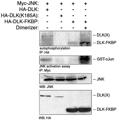
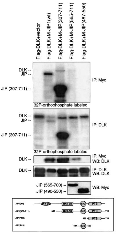
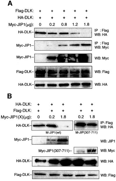
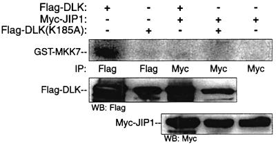
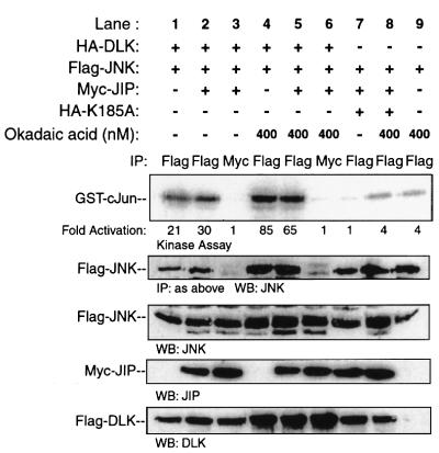
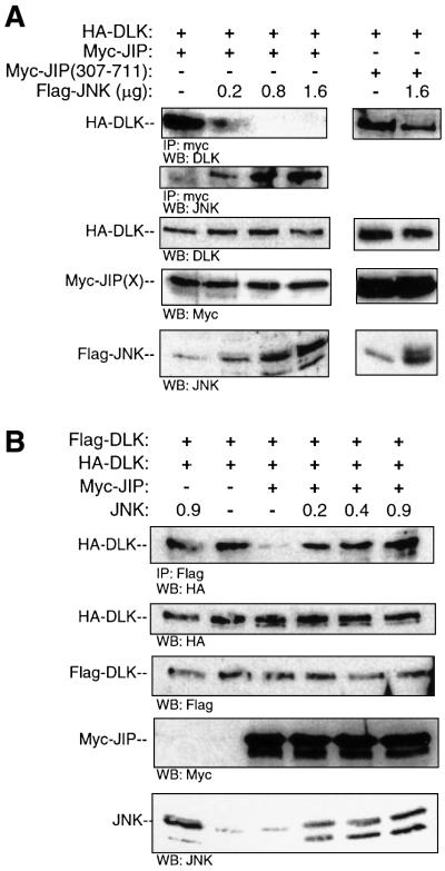
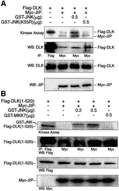
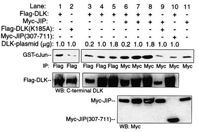
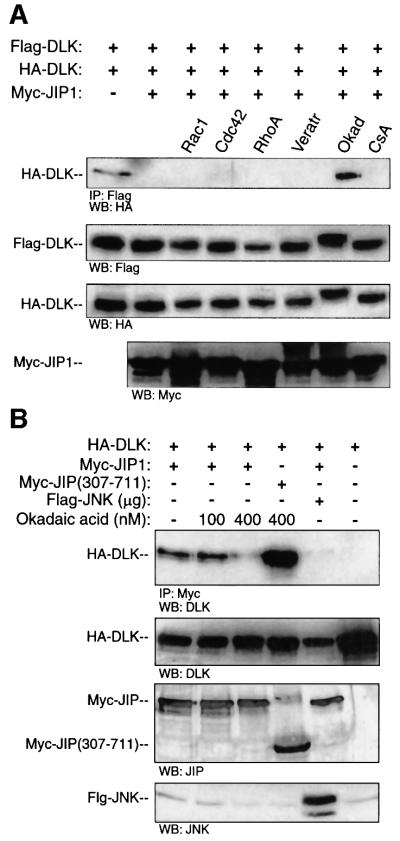
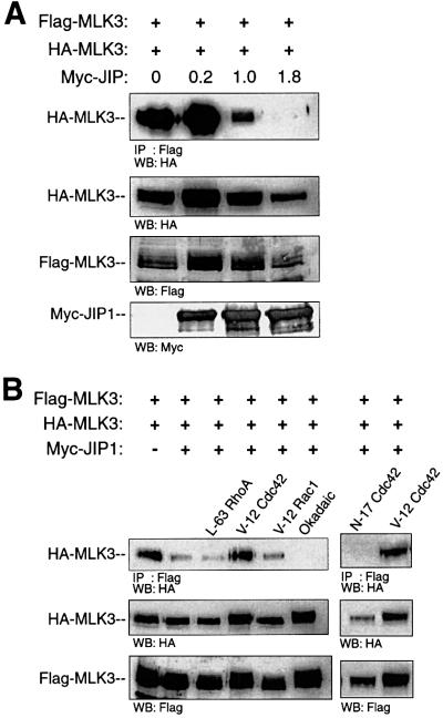
It has been proposed that JNK-interacting proteins (JIP) facilitate mixed lineage kinase-dependent signal transduction to JNK by aggregating the three components of a JNK module. A new model for the assembly and regulation of these modules is proposed based on several observations. First, artificially induced dimerization of dual leucine zipper-bearing kinase (DLK) confirmed that DLK dimerization is sufficient to induce DLK activation. Secondly, under basal conditions, DLK associated with JIP is held in a monomeric, unphosphorylated and catalytically inactive state. Thirdly, JNK recruitment to JIP coincided with significantly decreased affinity of JIP and DLK. JNK promoted the dimerization, phosphorylation and activation of JIP-associated DLK. Similarly, treatment of cells with okadaic acid inhibited DLK association with JIP and resulted in DLK dimerization in the presence of JIP. In summary, JIP maintains DLK in a monomeric, unphosphorylated, inactive state. Upon stimulation, JNK-JIP binding affinity increases while JIP-DLK interaction affinity is attenuated. Dissociation of DLK from JIP results in subsequent DLK dimerization, autophosphorylation and module activation. Evidence is provided that this model holds for other MLK-dependent JNK modules.


Figures

None
Fig. 1. DLK dimerization results in DLK autophosphorylation and JNK activation. COS 7 cells were transiently transfected with plasmid encoding Myc-JNK (0.5 µg) and with 0.5 µg of either HA-DLK, HA-DLK–FKBP or HA-DLK(K185A). After 24 h, dimerizer (5 nM) was added to the cells as indicated. DLK or its mutants were immunoprecipitated with HA antibodies, incubated in kinase buffer in the presence of [γ-32P]ATP, separated by SDS–PAGE and analyzed by autoradiography. JNK was immunoprecipitated from the same cell lysate and analyzed for catalytic activity. Cell lysates from corresponding experiments were immunoblotted using anti-JNK antibody to demonstrate equivalent expression of JNK. Anti-HA antibody was used to demonstrate the expression of DLK and its mutants.
None
Fig. 2. DLK associated with JIP is not phosphorylated. COS 7 cells were co-transfected as indicated with plasmids (0.2 µg each) encoding Flag-DLK, Myc-JIP1, Myc-JIP(307–711), Myc-JIP(565–700), Myc-JIP(490–550) and vector (to 2 µg), then metabolically labeled with [γ-32P]orthophosphate. DLK or JIP was immunoprecipitated as indicated. Immunoprecipitated complexes were separated by SDS–PAGE and analyzed by autoradiography. Corresponding cell lysates were immunoprecipitated with the indicated antibodies, separated by SDS–PAGE and analyzed by immunoblotting as indicated. Expression of wild-type-JIP1 and JIP(307–711) is seen in the top panel and expression of JIP(565–700) and JIP(490–550) is seen in the bottom panel. Experiments were repeated three times with similar results. M-, Myc.
None
Fig. 3. Wild-type JIP1 but not JIP(307–711) inhibits DLK dimerization. COS 7 cells were co-transfected with HA-DLK (0.1 µg), Flag-DLK (0.1 µg) and increasing quantities of Myc-JIP1 plasmids as indicated. (A) Flag-DLK complexes were immunoprecipitated from cell lysates using anti-Flag antibody, separated by SDS–PAGE and immunoblotted with either anti-HA or anti-Myc antibody. Correspond ing cell lysates were immunoblotted to evaluate the relative expression of Myc-JIP, HA-DLK and Flag-DLK proteins. (B) Cells were co-transfected with HA-DLK (0.1 µg), Flag-DLK (0.1 µg) and the indicated quantity of Myc-JIP or Myc-JIP(307–711) plasmids; samples were analyzed as above.
None
Fig. 4. JIP-associated DLK does not phosphorylate its specific substrate. COS 7 cells were co-transfected with plasmids encoding Flag-DLK (0.5 µg) or Flag-DLK(K185A) (0.5 µg) and Myc-JIP1 (0.2 µg) as indicated. Cell lysates were immunoprecipitated using the indicated antibodies. Immunoprecipitates were analyzed for DLK catalytic activity in vitro using GST–MKK7 as substrate. Immunoprecipitated complexes from corresponding experiments were separated by SDS–PAGE and were immunoblotted as indicated.
None
Fig. 5. Evaluation of the activation state of JIP-associated JNK expressed in cells. COS 7 cells were co-transfected as indicated with plasmids encoding Flag-JNK (0.5 µg), HA-DLK (0.2 µg), HA-DLK(K185A) (0.2 µg) and Myc-JIP1 (0.2 µg). At 24 h post-transfection, the indicated samples were treated for 3 h with 400 nM okadaic acid. Immunoprecipitation was performed with the indicated antibodies and immune complex-associated JNK was analyzed for catalytic activity using GST–c-Jun as substrate. Corresponding immunoprecipitated complexes were immunoblotted with anti-JNK antibody. Cell lysates from corresponding experiments were immunoblotted with the indicated antibodies to evaluate the expression of JNK, JIP and DLK. Fold activation is indicated relative to the control experiment shown in lane 7 for Flag immunoprecipitation experiments, and to the control experiment shown in lane 3 for Myc immunoprecipitation experiments.
None
Fig. 6. The presence of JNK results in decreased affinity of JIP1 for DLK and promotes DLK dimerization. (A) COS 7 cells were co-transfected with plasmids encoding HA-DLK (0.5 µg) and either Myc-JIP1 (0.5 µg) or Myc-JIP1(307–711) (0.5 µg) and increasing quantities of plasmid encoding Flag-JNK as indicated. Cell lysates were immunoprecipitated with anti-Myc antibody, separated on SDS–PAGE and immunoblotted with anti-DLK antibody. Cell lysates from corresponding experiments were immunoblotted with the indicated antibodies to evaluate the expression of JNK, JIP and DLK. (B) COS 7 cells were co-transfected as indicated with HA-DLK (0.05 µg), Flag-DLK (0.05 µg), Myc-JIP1 (0.9 µg), the indicated quantity of JNK and vector (to 2 µg). Cell lysates were immuno precipitated with anti-Flag antibody and immunoblotted with HA antibody. Corresponding cell lysates were immunoblotted with the indicated antibodies to evaluate the expression of HA-DLK, Flag-DLK, Myc-JIP1 and JNK.
None
Fig. 7. JNK promotes JIP-associated DLK phosphorylation in vitro. COS 7 cells were co-transfected as indicated with plasmids encoding the Flag-DLK (A) or Flag-DLK(1–520) (0.5 µg) (B) and Myc-JIP1 (0.5 µg). Cell lysates were immunoprecipitated using either anti-Flag or anti-Myc antibody as indicated. Immunoprecipitated complexes were analyzed in vitro for DLK phosphorylation by incubating them in kinase buffer containing [γ-32P]ATP and the indicated quantity of GST–JNK, kinase-dead GST–JNK(K55R) or GST–MKK7. Immunoprecipitated complexes were separated by SDS–PAGE and analyzed by autoradiography. Alternatively, immunoprecipitated complexes were immunoblotted with anti-Flag antibody. Expression of proteins was assessed by immunoblotting corresponding cell lysates.
None
Fig. 8. JIP N-terminus is required for JNK activation by JIP-associated DLK in vitro. Myc-JIP1 (0.2 µg), Myc-JIP1(307–711) (0.2 µg) or vector control were co-transfected with either inactive Flag-DLK(K185A) or various quantities of Flag-DLK plasmid. DLK, DLK(K185A) or JIP1 was immunoprecipitated from the cell lysates using the indicated antibodies. Immunoprecipitates were then assayed in vitro for their ability to activate GST–JNK. Kinase buffer containing recombinant GST–MKK7, GST–JNK, GST–c-Jun and [γ-32P]ATP was added to immunoprecipitated complexes and incubated at 30°C for 15 min. Immunoprecipitates were separated on SDS–PAGE, transferred to nitrocellulose and autoradiographed. Immunoblots from corresponding cell lysates were used to evaluate the relative expression of DLK and JIP in each reaction. Similar results were obtained in three independent experiments.
None
Fig. 9. Okadaic acid treatment results in decreased affinity of JIP for DLK and DLK dimerization in the presence of JIP. (A) COS 7 cells were transfected with plasmids encoding HA-DLK (0.1 µg), Flag-DLK (0.1 µg) and Myc-JIP1 (1.0 µg) as indicated. The indicated cells were transfected simultaneously with activated forms of either Myc-V12Cdc42Hs, Myc-V12Rac1 or Myc-L63RhoA. Where indicated, cells were treated for 3 h with either veratridine (20 µg), okadaic acid (400 nM) or cyclosporin A (30 µM). Flag-DLK was immuno precipitated from cell lysates; immunoprecipitated complexes were separated by SDS–PAGE and were immunoblotted with anti-HA antibody. Immunoblots from the corresponding cell lysates were used to evaluate the relative expression of Flag-DLK, HA-DLK and Myc-JIP. (B) COS 7 cells were co-transfected with plasmids encoding HA-DLK (0.5 µg) and Myc-JIP1 (0.5 µg). Cells were treated for 3 h with the indicated concentrations of okadaic acid. Cell lysates were immunoprecipitated with anti-Myc antibody, separated by SDS–PAGE and immunoblotted with anti-DLK antibody. Cell lysates from the corresponding experiments were immunoblotted with the indicated antibodies to evaluate the expression of JIP and DLK. These experiments were repeated three times with similar results.
None
Fig. 10. JIP inhibits dimerization of MLK3 and V12Cdc42Hs promotes MLK3 dimerization in the presence of JIP. (A) COS 7 cells were co-transfected with Flag-MLK3 (0.1 µg), HA-MLK3 (0.1 µg) and increasing quantities of Myc-JIP plasmids as indicated. Flag-MLK3 complexes were immunoprecipitated from cell lysates using anti-Flag antibody, separated by SDS–PAGE and immunoblotted with anti-HA antibody. Corresponding cell lysates were immunoblotted to evaluate the relative expression of Myc-JIP, HA-MLK3 and Flag-MLK3 proteins. (B) COS 7 cells were transfected with Flag-MLK3 (0.05 µg), HA-MLK3 (0.05 µg), Myc-JIP (1.0 µg) and either V12Rac1, V12Cdc42Hs or L63RhoA plasmids as indicated. Where indicated, 24 h after transfection, cells were treated for 3 h with okadaic acid (400 nM). As above, immunoprecipitation and immunoblotting were used to assess the association of HA-MLK3 and Flag-MLK3 in the presence of JIP. Experiments were repeated three times with similar results.

Similar articles

Cited by

References

    1. Barancik M., Htun,P. and Schaper,W. (1999) Okadaic acid and anisomycin are protective and stimulate the SAPK/JNK pathway. J. Cardiovasc. Pharmacol., 34, 182–190. - PubMed
    1. Bock B.C., Vacratsis,P.O., Qamirani,E. and Gallo,K.A. (2000) Cdc42-induced activation of the mixed-lineage kinase SPRK in vivo. Requirement of the Cdc42/Rac interactive binding motif and changes in phosphorylation. J. Biol. Chem., 275, 14231–14241. - PubMed
    1. Burack R.W. and Shaw,A.S. (2000) Signal transduction: hanging on scaffold. Curr. Opin. Cell Biol., 12, 211–216. - PubMed
    1. Charest D.L., Mordet,G., Harder,K.W., Jirik,F. and Pelech,S.L. (1993) Molecular cloning, expression and characterization of the human mitogen-activated protein kinase p44erk1. Mol. Cell. Biol., 13, 4679–4690. - PMC - PubMed
    1. Chen Y.R., Meyer,C.F. and Tan,T.H. (1996) Persistent activation of c-Jun N-terminal kinase 1 (JNK1) in γ radiation-induced apoptosis. J. Biol. Chem., 271, 631–634. - PubMed

Publication types

MeSH terms

Substances